Review
. 2019 Jun;35(Suppl 2):47-56.
doi: 10.1007/s12055-019-00832-z. Epub 2019 May 30.

Aortitis and aortic aneurysm in systemic vasculitis

Affiliations
Review

Aortitis and aortic aneurysm in systemic vasculitis

Sanjay Tyagi et al. Indian J Thorac Cardiovasc Surg. 2019 Jun.

Abstract

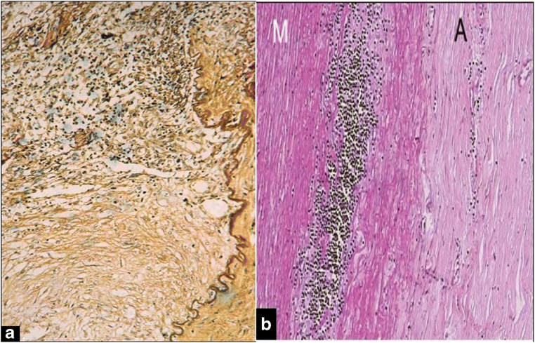
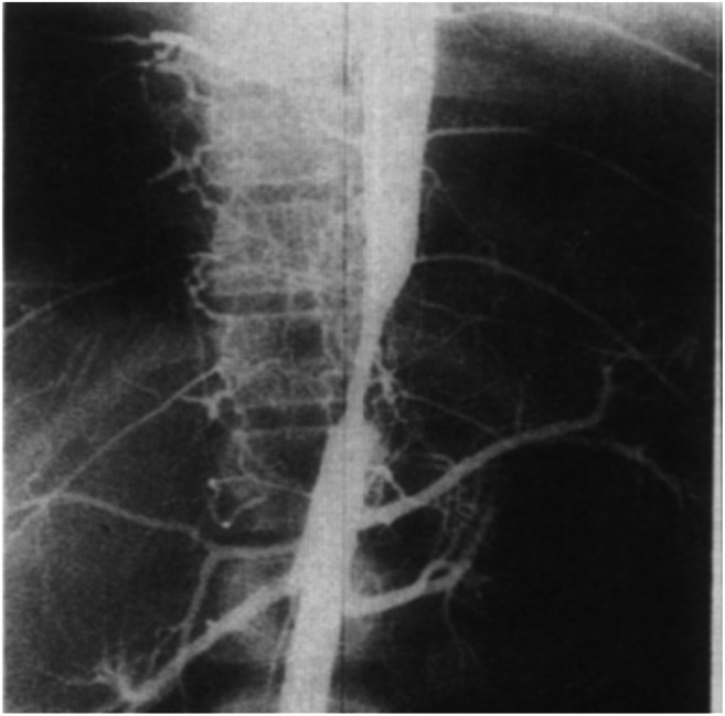
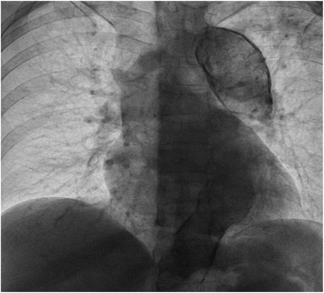
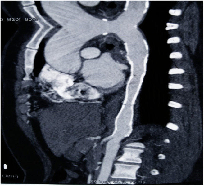
The term "large-vessel vasculitis" refers to chronic inflammation affecting the elastic arteries, mainly aorta and its major branches. Large-vessel vasculitis affecting the aorta is termed as "aortitis." This leads to diffuse wall thickening, loss of elasticity, stenosis, occlusion, dissection, calcification or aneurysm formation. Vasculitis involving the aorta includes Takayasu arteritis (commonest large-vessel vasculitis in India), giant cell arteritis (GCA), the periaortitis spectrum (including inflammatory abdominal aortic aneurysm), rheumatic diseases and IgG4-related disease. It is important for physicians to be aware of the aortic manifestations of common vasculitis-so that clinically relevant aortic involvement is not missed. Also, it is equally important for surgeons to keep in mind that aortic aneurysms can have an inflammatory aetiology. The primary reason for this is the central role that immunosuppressive therapy plays in the management of inflammatory aortic aneurysms. Surgical or percutaneous interventions, when needed, are most likely to succeed when performed after activity control; perioperative steroids may be beneficial in select cases, and postoperative disease activity control is imperative.

Keywords: Aortic aneurysm; Aortitis; Giant cell arteritis; Takayasu arteritis.


Conflict of interest statement

Conflicts of interestThe authors declare they have no conflict of interest.

Figures

Fig. 1
Fig. 1
a Granulomatous inflammation of descending thoracic aorta involving junction of tunica media and adventitia during active phase of TA. b Mononuclear cell infiltration of tunica media (M) and fibrosis in adventitia (A) during chronic phase of TA [images from Vaideeswar and Deshpande [8] with permission]
Fig. 2
Fig. 2
a Multi-focal aortic disease with skip areas showing a large aneurysm in the abdominal aorta. The wall of the aneurysm when studied histologically also showed the presence of chronic dissection as seen in b haematoxylin and eosin (×100) and c elastic van Gieson (×100). Arrow points to the intimal flap. The wall is represented only by thickened intima (I) and adventitia (A) [images from Vaideeswar and Deshpande [8] with permission]
Fig. 3
Fig. 3
Aortogram shows severe stenosis of descending thoracic aorta in a young girl with TA. Patient had hypertension and congestive heart failure, which were relieved after balloon angioplasty
Fig. 4
Fig. 4
Aortogram of abdominal aorta shows occlusion of infrarenal aorta in a patient with TA
Fig. 5
Fig. 5
Fluoroscopy shows marked calcification with diffuse dilatation of descending thoracic aorta in a young hypertensive female with TA. There was no stenotic involvement of the aorta
Fig. 6
Fig. 6
CT scan shows aneurysmal dilatation of root, ascending aorta and innominate artery in a patient of TA who had previously been stented (white borders) for descending aorta and left subclavian artery stenosis
Fig. 7
Fig. 7
DSA of arch of aorta in a young female with Takayasu’s arteritis with aneurysmal involvement of left carotid and subclavian arteries (in addition to stenotic involvement of all arch vessels)
Fig. 8
Fig. 8
Computed tomography aortogram (CTA) showing a aortic dissection (Stanford type B) of the descending thoracic aorta leading to severe narrowing of true lumen of aorta by displaced intimal flap (white arrow). Yellow circle denotes the proximal landing zone of the stent graft; here, the diameter of the aorta is 13.6 mm. b Three-dimensional (3D) reconstructed image showing aortic dissection. c Three-dimensional reconstructed image showing the completely sealed-off aortic dissection and well-expanded stent graft. d CTA at 1-year follow-up showing good expansion of stent graft and true lumen (figure from Tyagi et al [22])

References

    1. Caspary L. Inflammatory diseases of the aorta. Vasa. 2016;45:17–29. doi: 10.1024/0301-1526/a000491. - DOI - PubMed
    1. Töpel I, Zorger N, Steinbauer M. Inflammatory diseases of the aorta. Part 1: non-infectious aortitis. Gefässchirurgie. 2016;21:S80–S86. doi: 10.1007/s00772-016-0143-9. - DOI - PMC - PubMed
    1. Loricera J, Blanco R, Hernández JL, et al. Non-infectious aortitis: a report of 32 cases from a single tertiary centre in a 4-year period and literature review. Clin Exp Rheumatol. 2015;33:S–19-31. - PubMed
    1. Miller DV, Isotalo PA, Weyand CM, Edwards WD, Aubry MC, Tazelaar HD. Surgical pathology of noninfectious ascending aortitis: a study of 45 cases with emphasis on an isolated variant. Am J Surg Pathol. 2006;30:1150–1158. doi: 10.1097/01.pas.0000213293.04026.ec. - DOI - PubMed
    1. Pacini D, Leone O, Turci S, et al. Incidence, etiology, histologic findings, and course of thoracic inflammatory aortopathies. Ann Thorac Surg. 2008;86:1518–1523. doi: 10.1016/j.athoracsur.2008.07.039. - DOI - PubMed

LinkOut - more resources