. 2020 Sep 30:13:9623-9629.
doi: 10.2147/OTT.S263250. eCollection 2020.

The Impact of Smoking on Pulmonary Metastasis in Colorectal Cancer

Affiliations

The Impact of Smoking on Pulmonary Metastasis in Colorectal Cancer

Akitsugu Makino et al. Onco Targets Ther. .

Abstract

Introduction: Recently, clinical studies have revealed that smoking can contribute to the poor prognosis of colorectal cancer (CRC) and, additionally, can be a risk factor for pulmonary metastasis of CRC. However, there has been no basic research regarding the underlying molecular mechanism. The purpose of this study was to clarify the mechanism by which smoking causes pulmonary metastasis of CRC.

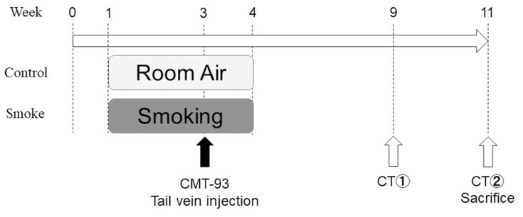
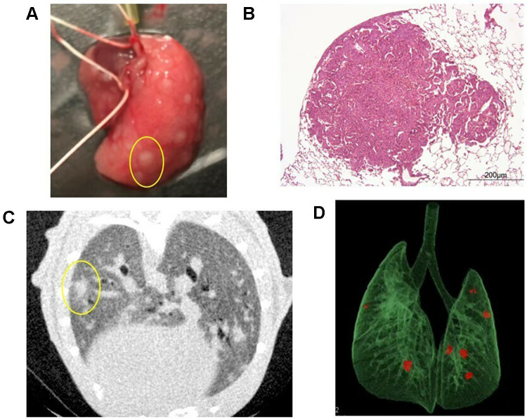
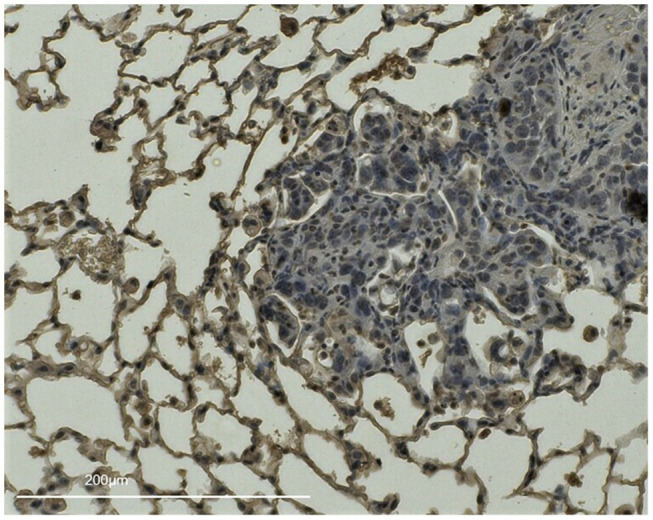
Methods: First, pulmonary metastasis model mice inhaled cigarette smoke or air (control) for 1 h once a day for 3 weeks. We attempted to clarify the effect of smoking on the incidence of pulmonary metastasis. On the 15th day, CMT-93 cells were injected into the tail vein. At 6 and 8 weeks following injection, the extent of pulmonary metastasis was evaluated using in vivo micro CT. After the last CT examination, the mice were sacrificed, and the lungs were extracted for pathological examination.

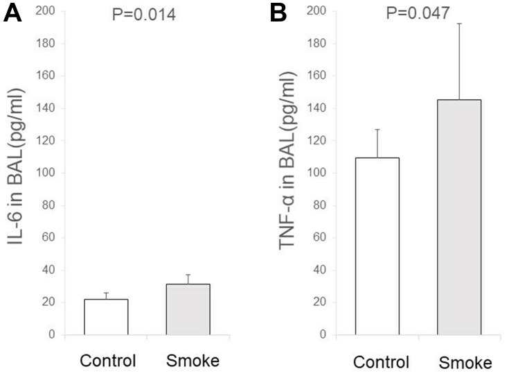
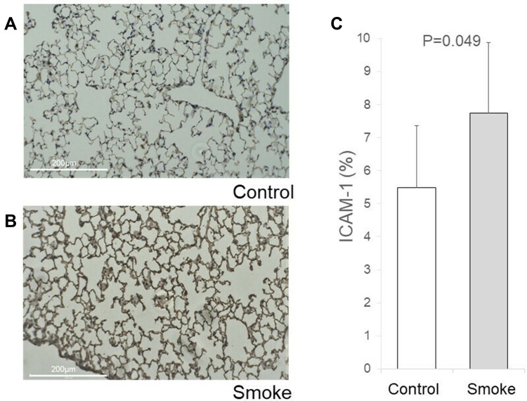
Results: The number of mice with pulmonary metastases in the smoking group was significantly higher than in the control group. Three weeks of smoking induced mild inflammation in the lungs, as evidenced by increases in the levels of IL-6 and TNF-α in bronchoalveolar lavage. Moreover, the adhesion-related molecule ICAM-1 was overexpressed in pulmonary tissue, which allowed drained cancer cells to remain in the lung and contribute to the formation of pulmonary metastasis.

Conclusion: Collectively, cigarette smoking may contribute to the pathogenesis and development of pulmonary metastasis in CRC through enhancement of adhesion and inflammation.

Keywords: ICAM-1; adhesion molecules; colorectal cancer; pulmonary metastasis; smoking.


Conflict of interest statement

Prof. Dr. Yuko Kitagawa reports grants and/or personal fees from ASAHI KASEI PHARMA CO., LTD., TAIHO PHARMACEUTICAL CO., LTD, CHUGAI PHARMACEUTICAL CO., LTD., DAIICHI SANKYO COMPANY, LIMITED, Merck Serono Co., Ltd., EA Pharma Co., Ltd., Yakult Honsha Co. Ltd., Otsuka Pharmaceutical Co., Ltd., Takeda Pharmaceutical Co., Ltd., Otsuka Pharmaceutical Factory Inc., SHIONOGI & CO., LTD., KAKEN PHARMACEUTICAL CO., LTD., Kowa Pharmaceutical Co., Ltd., Astellas Pharma Inc., MEDICON INC., DAINIPPON SUMITOMO PHARMA Co., Ltd., Taisho Toyama Pharmaceutical Co., Ltd., Kyowa Hakko Kirin Co., Ltd., Pfizer Japan Inc., ONO PHARMACEUTICAL CO., LTD., NIHON PHARMACEUTICAL CO., LTD., Japan Blood Products Organization, Medtronic Japan Co., Ltd., Sanofi K.K., Eisai Co., Ltd., TSUMURA & CO., KCI Licensing, Inc., ABBOTT JAPAN CO., LTD., FUJIFILM Toyama Chemical Co., Ltd., outside the submitted work. The authors report no other conflicts of interest in this work.

Figures

Figure 1
Figure 1
Animal experimental protocol, The smoking group mice were exposed to smoke for 60 min/day on weekdays for 3 weeks. The control group mice were exposed to air over the same time period. On the 15th day of smoking exposure, 3.5 × 10⁶ CMT-93 cells were injected into the tail vein. Six and eight weeks following injection, the extent of pulmonary metastasis was evaluated using in vivo micro CT. All mice were sacrificed immediately after the last CT examination and the lungs were extracted.
Figure 2
Figure 2
Pulmonary metastases. (A) Macroscopic findings of pulmonary metastasis in the right lobe of the lung. In this mouse, many nodules were identified (one is circled in yellow). (B) Hematoxylin-eosin staining of pulmonary metastasis in a mouse. (C) A representative two-dimensional image of a micro CT scan in a smoking mouse. In this mouse, one nodule was identified in the left lung (circled in yellow). (D) CT reconstruction of a pulmonary metastasis from a mouse model. Three-dimensional microstructural image data were reconstructed using Tri/3D-BON software. Red colored nodules indicate pulmonary metastasis.
Figure 3
Figure 3
IL-6 and TNF-α in the bronchoalveolar lavage (BAL). The protein levels of IL-6 and TNF-α in the BAL were measured. The BAL was collected from the left lobe by washing three times with 500 μL saline through the left bronchus. The concentrations of IL-6 and TNF-α in the BAL were determined using a mouse IL-6 Quantikine ELISA kit and a mouse TNF-α ELISA Development Kit. (A) IL-6 expression in the BAL was significantly higher in the smoking group compared with the control group (31.2 ± 6.2 vs 22.1 ± 3.8, P = 0.014). (B) TNF-α expression in BAL was significantly higher in the smoking group compared with the control group (145.5 ± 46.7 vs 109.5 ± 17.5, P = 0.047).
Figure 4
Figure 4
Immunohistochemical (IHC) staining of ICAM-1. Lung tissues from the largest longitudinal section in the right lobe of control and smoking mice were stained for ICAM-1. Anti-ICAM1 primary antibody diluted 1:100 was used for IHC staining. ICAM-1 expression was quantified by measuring the percentage area of expression (0–100%) using ImageJ. Briefly, five randomly chosen field images (0.02 × 0.01 inches) from the slide containing the maximum longitudinal section of the right lobe were acquired using a light microscope, and colors that were not of interest were removed via the replace mode. The adapted images were converted to grey scale and the area of expression was located by adjusting the threshold. The percentage area of expression is denoted by positive pixels on the labeled areas. (A) IHC staining of ICAM-1 in control mouse lung. (B) IHC staining of ICAM-1 in smoking mouse lung. (C) The ICAM-1 antibody tended to strongly stain the lungs from the smoking group (7.75 ± 3.27 vs 5.48 ± 2.1, P = 0.049).
Figure 5
Figure 5
IHC staining of ICAM-1 in a smoking mouse lung with pulmonary metastasis, Lung tissues from the largest longitudinal section in the right lobe of a smoking group mouse injected with CMT-93 cells on the 15th day of smoking exposure were stained for ICAM-1. There are some pulmonary metastases surrounded by areas enhanced with ICAM-1.

Similar articles

Cited by

References

    1. Sobus SL, Warren GW. The biologic effects of cigarette smoke on cancer cells. Cancer. 2014;120(23):3617–3626. doi:10.1002/cncr.28904 - DOI - PubMed
    1. Hackshaw AK, Law MR, Wald NJ. The accumulated evidence on lung cancer and environmental tobacco smoke. BMJ. 1997;315(7114):980–988. doi:10.1136/bmj.315.7114.980 - DOI - PMC - PubMed
    1. Malhotra J, Borron C, Freedman ND, et al. Association between Cigar or Pipe Smoking and Cancer Risk in Men: A Pooled Analysis of Five Cohort Studies. Cancer Prevent Res. 2017;10(12):704–709. - PubMed
    1. Kuang JJ, Jiang ZM, Chen YX, et al. Smoking Exposure and Survival of Patients with Esophagus Cancer: A Systematic Review and Meta-Analysis. Gastroenterol Res Pract. 2016;2016:7682387. - PMC - PubMed
    1. Inoue-Choi M, Hartge P, Liao LM, Caporaso N, Freedman ND. Association between long-term low-intensity cigarette smoking and incidence of smoking-related cancer in the national institutes of health-AARP cohort. Int j Cancer. 2018;142(2):271–280. - PMC - PubMed