

Dermatological Manifestations in the Intensive Care Unit: A Practical Approach
- PMID: 33062328
- PMCID: PMC7533796
- DOI: 10.1155/2020/9729814
Dermatological Manifestations in the Intensive Care Unit: A Practical Approach
Abstract
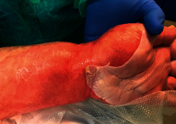
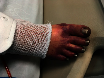
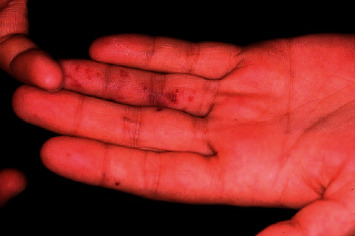
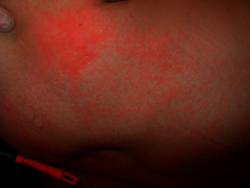
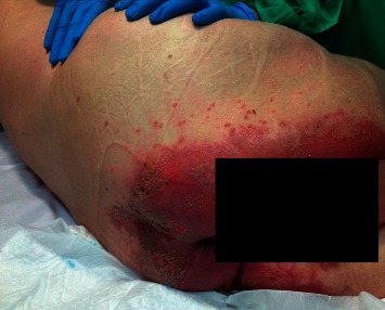
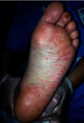
Dermatological problems are not usually related to intensive medicine because they are considered to have a low impact on the evolution of critical patients. Despite this, dermatological manifestations (DMs) are relatively frequent in critically ill patients. In rare cases, DMs will be the main diagnosis and will require intensive treatment due to acute skin failure. In contrast, DMs can be a reflection of underlying systemic diseases, and their identification may be key to their diagnosis. On other occasions, DMs are lesions that appear in the evolution of critical patients and are due to factors derived from the stay or intensive treatment. Lastly, DMs can accompany patients and must be taken into account in the comprehensive pathology management. Several factors must be considered when addressing DMs: on the one hand, the moment of appearance, morphology, location, and associated treatment and, on the other hand, aetiopathogenesis and classification of the cutaneous lesion. DMs can be classified into 4 groups: life-threatening DMs (uncommon but compromise the patient's life); DMs associated with systemic diseases where skin lesions accompany the pathology that requires admission to the intensive care unit (ICU); DMs secondary to the management of the critical patient that considers the cutaneous manifestations that appear in the evolution mainly of infectious or allergic origin; and DMs previously present in the patient and unrelated to the critical process. This review provides a characterization of DMs in ICU patients to establish a better identification and classification and to understand their interrelation with critical illnesses.
Copyright © 2020 Mariona Badia et al.
Conflict of interest statement
The authors declare that they have no conflicts of interest.
Figures
References
-
- George S. M., Harrison D. A., Welch C. A., Nolan K. M., Friedmann P. S. Dermatological conditions in intensive care: a secondary analysis of the intensive care national audit & research centre (ICNARC) case mix programme database. Critical Care. 2008;12(1):p. S1. doi: 10.1186/cc6141. https://www.ncbi.nlm.nih.gov/pmc/articles/PMC2607109/ - DOI - PMC - PubMed
-
- Agrawal P., Peter J. V., George R. Dermatological manifestations and relationship to outcomes of patients admitted to a medical intensive care unit: a study from a tertiary care hospital in India. Postgraduate Medical Journal. 2013;89(1055):501–507. doi: 10.1136/postgradmedj-2012-131610. - DOI - PubMed
Publication types
LinkOut - more resources
Full Text Sources
