

Symptomatic Cavum Septum Pellucidum Cyst: A Rare Presentation
- PMID: 33062515
- PMCID: PMC7550031
- DOI: 10.7759/cureus.10395
Symptomatic Cavum Septum Pellucidum Cyst: A Rare Presentation
Abstract
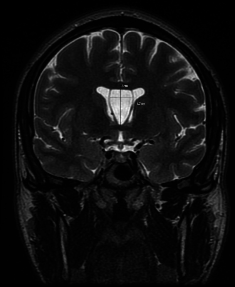
A cavum septum pellucidum is a cerebrospinal fluid (CSF) filled cavity situated between the lateral ventricles and is considered as a normal anatomic variant sporadically seen on neuroimaging. While a cavum septum pellucidum is a relatively uncommon incidental neuroimaging finding, symptomatic cysts of the cavum septum pellucidum are very rare, with only a few cases reported in the literature so far. They are defined as fluid-filled structures with lateral bowing of the walls and membranes separated by at least 10 mm or more. We present the case of a 25-year-old male patient with a rapidly expanding cyst of the septum pellucidum with headaches refractory to conventional pharmacological therapy. A 3T magnetic resonance imaging (MRI) of the brain with contrast was performed, which confirmed the diagnosis. Due to the failure of non-interventional treatment, he was treated with therapeutic endoscopic fenestration of the cyst. Postoperatively, he reported a complete resolution of the presenting symptoms.
Keywords: cavum septum pellucidum; csf fluid dynamics; cyst fenestration; endoscopic repair; headache; hydrocephalus; intracranial cyst; neuroendoscopy; positional headache; refractory headache.
Copyright © 2020, Pillai et al.
Conflict of interest statement
The authors have declared that no competing interests exist.
Figures

References
-
- Headache profiles in patients with a dilatated cyst of the cavum septi pellucidi. Wang KC, Fuh JL, Lirng JF, Huang WC, Wang SJ. Cephalalgia. 2004;24:867–874. - PubMed
-
- The septum pellucidum: normal and abnormal. Sarwar M. https://pubmed.ncbi.nlm.nih.gov/2505543/ AJNR Am J Neuroradiol. 1989;10:989–1005. - PMC - PubMed
-
- Incidence of cavum septi pellucidi and cavum vergae in 1,032 human brains. Schwidde JT. AMA Arch Neurol Psychiatry. 1952;67:625–632. - PubMed
-
- Cavum septi pellucidi and cavum vergae | MedLink Neurology. [Dec;2018 ];Barth P, Siebert J. https://www.medlink.com/article/cavum_septi_pellucidi_and_cavum_vergae Medlink Neurology. 1995
Publication types
LinkOut - more resources
Full Text Sources