. 2021 Feb;21(2):525-539.
doi: 10.1111/ajt.16358. Epub 2020 Nov 3.

Mus musculus papillomavirus 1 is a key driver of skin cancer development upon immunosuppression

Affiliations

Mus musculus papillomavirus 1 is a key driver of skin cancer development upon immunosuppression

Sonja Dorfer et al. Am J Transplant. 2021 Feb.

Abstract

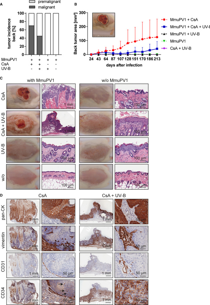
Epidemiological and experimental data implicate cutaneous human papillomavirus infection as co-factor in the development of cutaneous squamous cell carcinomas (cSCCs), particularly in immunocompromised organ transplant recipients (OTRs). Herein, we established and characterized a skin cancer model, in which Mus musculus papillomavirus 1 (MmuPV1) infection caused cSCCs in cyclosporine A (CsA)-treated mice, even in the absence of UV light. Development of cSCCs and their precursors were observed in 70% of MmuPV1-infected, CsA-treated mice on back as well as on tail skin. Immunosuppression by systemic CsA, but not UV-B irradiation, was a prerequisite, as immunocompetent or UV-B-irradiated mice did not develop skin malignancies after infection. In the virus-driven cSCCs the MmuPV1-E6/E7 oncogenes were abundantly expressed, and transcriptional activity and productive infection demonstrated. MmuPV1 infection induced the expression of phosphorylated H2AX, but not degradation of proapoptotic BAK in the cSCCs. Transfer of primary cells, established from a MmuPV1-induced cSCC from back skin, into athymic nude mice gave rise to secondary cSCCs, which lacked viral DNA, demonstrating that maintenance of the malignant phenotype was virus independent. This papillomavirus-induced skin cancer model opens future investigations into viral involvement, pathogenesis, and cancer surveillance, aiming at understanding and controlling the high incidence of skin cancer in OTRs.

Keywords: animal models: murine; basic (laboratory) research / science; cancer / malignancy / neoplasia: skin - nonmelanoma; dermatology; immunosuppressant - calcineurin inhibitor: cyclosporine A (CsA); immunosuppression / immune modulation; infection and infectious agents - viral; infectious disease.


Conflict of interest statement

The authors of this manuscript have no conflicts of interest to disclose as described by the American Journal of Transplantation.

Figures

FIGURE 1
FIGURE 1
Tumor incidence after experimental MmuPV1 skin infection. (A) Tumor incidence on back skin in MmuPV1‐infected mice at week 30 postinfection. None of the uninfected mice developed skin tumors. (B) Time course of tumor outgrowth on back skin. Tumor area is given in mm2. (C) Representative mouse of each experimental group with corresponding HE image. Left panel, MmuPV1‐infected mice; right panel, uninfected mice. (D) Pan‐cytokeratin, vimentin, CD31, and CD34 stainings of cSCCs of back skin. One representative MmuPV1‐infected, CsA‐treated (far‐left and left panels) and MmuPV1‐infected CsA‐/UV‐B–treated (far‐right and right panels) mouse is depicted. Overview and higher magnification of the same tumors are shown
FIGURE 2
FIGURE 2
Viral presence in tumors on back skin. (A) Left panel: MmuPV1 genome copy numbers in back skin of MmuPV1‐infected mice stratified according to tumor development. Right panel: MmuPV1‐E1^E4 spliced transcripts in back skin tumors and nontumorous back skin of infected mice. (B) Left panel: MmuPV1‐E6/E7 mRNA in back skin of infected mice. Middle panel: IHC for MmuPV1‐L1/L2 capsid proteins in back skin. Right panel: corresponding HE‐stained sections
FIGURE 3
FIGURE 3
yH2AX and CPD staining of back skin. (A) Representative yH2AX (far‐left panel) and CPD (left panel) stainings of MmuPV1‐infected mice. Representative yH2AX (right panel) and CPD (far‐right panel) stainings of uninfected control mice. (B) Quantification of yH2AX‐immunopositive back skin area. (C) Quantification of CPD‐immunopositive back skin area
FIGURE 4
FIGURE 4
BAK staining of back skin. (A) Representative BAK stainings of MmuPV1‐infected (left panel) and uninfected (right panel) mice. (B) Quantification of BAK‐immunopositive back skin area. (C) Quantification of BAK immunostainings in tumorous and nontumorous back skin of MmuPV1‐infected, CsA‐treated and MmuPV1‐infected, CsA‐/UV‐B–treated mice
FIGURE 5
FIGURE 5
Presence of CD4+ and CD8+ T cells in back skin. (A) Representative CD4+ (far‐left panel) and CD8+ (left panel) stainings of MmuPV1‐infected mice. Representative CD4+ (right panel) and CD8+ (far‐right panel) stainings of uninfected control mice. (B) Quantification of CD4+‐immunopositive T cells per mm2 back skin in back skin. (C) Quantification of CD8+‐immunopositive T cells per mm2 back skin. (D) Quantification of CD4+‐immunopositive T cells in tumorous and nontumorous back skin of MmuPV1‐infected, CsA‐treated and MmuPV1‐infected, CsA‐/UV‐B–treated mice. (E) Quantification of CD8+‐immunopositive T cells in tumorous and nontumorous back skin of MmuPV1‐infected, CsA‐treated and MmuPV1‐infected, CsA‐/UV‐B–treated mice
FIGURE 6
FIGURE 6
Presence of CD4+ and CD8+ T cells in draining lymph nodes. (A) CD4+ stainings of lymph nodes derived from MmuPV1‐infected mice (far‐left and left panels). CD4+ stainings of lymph nodes derived from uninfected mice (far‐right and right panels). Overview and higher magnification of the same tumors are shown. (B) Quantification of CD4+‐immunopositive lymph node areas. (C) CD8+ stainings of lymph nodes derived from MmuPV1‐infected mice (far‐left and left panels). CD8+ stainings of lymph nodes derived from uninfected mice (far‐right and right panels). Overview and higher magnification of the same tumors are shown. (D) Quantification of CD8+‐immunopositive lymph node areas
FIGURE 7
FIGURE 7
Presence of CD45R+ T cells in draining lymph nodes. (A) CD45R+ stainings of lymph nodes derived from MmuPV1‐infected mice (far‐left and left panels). CD45R+ stainings of lymph nodes derived from uninfected mice (far‐right and right panels). Overview and higher magnification of the same tumors are shown. (B) Quantification of CD45R+‐immunopositive lymph node areas
FIGURE 8
FIGURE 8
MmuPV1‐specific neutralizing antibodies in mouse sera. IC50 titers were determined by PsV‐NA
FIGURE 9
FIGURE 9
Tumor cell explant and virus‐independent secondary tumor formation. (A) Primary tumor cells established from a MmuPV1‐infected, CsA‐/UV‐B–treated mouse. (B) Intradermal administration of primary cSCC cells of passage 11 to NMRI‐Foxn1nu/nu mice gave rise to secondary tumors. One representative NMRI‐Foxn1nu/nu mouse 30 days postinjection. Secondary tumors after procurement. Overview and higher magnification of a HE‐stained secondary cSCC. (C) Presence of MmuPV1 DNA in the primary cSCC and in the early passages of the primary cSCC cell line. Absence of MmuPV1 DNA in late passages of the primary cSCC cell line and in the secondary tumor

References

    1. Karia PS, Han J, Schmults CD. Cutaneous squamous cell carcinoma: estimated incidence of disease, nodal metastasis, and deaths from disease in the United States, 2012. J Am Acad Dermatol. 2013;68(6):957–966. - PubMed
    1. Rogers HW, Weinstock MA, Feldman SR, et al. Incidence estimate of nonmelanoma skin cancer (keratinocyte carcinomas) in the U.S. population, 2012. JAMA Dermatology. 2015;151(10):1081–1086. - PubMed
    1. Howley PM, Pfister HJ. Beta genus papillomaviruses and skin cancer. Virology. 2015;479–480:290–296. - PMC - PubMed
    1. Garrett GL, Blanc PD, Boscardin J, et al. Incidence of and risk factors for skin cancer in organ transplant recipients in the United States. JAMA Dermatology. 2017;153(3):296–303. - PubMed
    1. Mudigonda T, Levender MM, O'Neill JL, et al. Incidence, risk factors, and preventative management of skin cancers in organ transplant recipients: a review of single‐ and multicenter retrospective studies from 2006 to 2010. Dermatol Surg. 2013;39(3 Pt 1):345–364. - PubMed

Publication types