

[Preliminary exploration on the application of hydrogel from acellular porcine adipose tissue to assist lipofilling]
- PMID: 33063500
- PMCID: PMC8171868
- DOI: 10.7507/1002-1892.202002126
[Preliminary exploration on the application of hydrogel from acellular porcine adipose tissue to assist lipofilling]
Abstract
Objective: To investigate the effect of hydrogel from acellular porcine adipose tissue (HAPA) on the survival of transplanted adipose tissue.
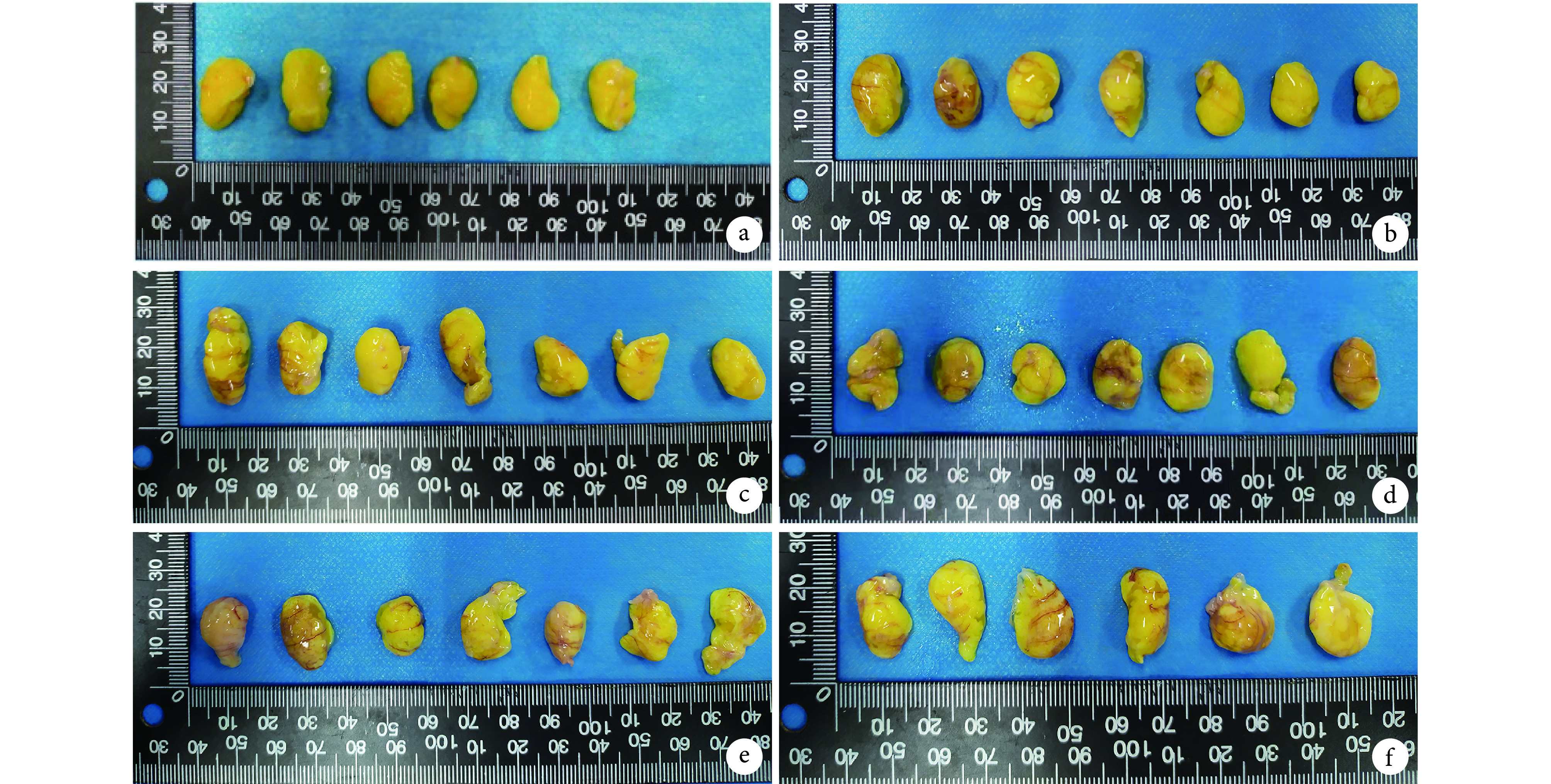
Methods: For in vitro study, adipose tissue and HAPA-adipose tissue complex were cultured in normoxia and hypoxia atmospheres for 24 and 72 hours. TUNEL and Perilipin immunofluorescence staining were performed to observe the effect of HAPA on apoptosis and survival of adipocities. For in vivo study, 42 healthy male nude mice (4-6 weeks old) weighing 15-18 g were randomly divided into adipose group (group A), 10%HAPA group (group B), 20%HAPA group (group C), 30%HAPA group (group D), 40%HAPA group (group E), and 50%HAPA group (group F) according to different HAPA/adipose tissue volume ratio ( n=7). For each group, 1 mL adipose tissue or HAPA-adipose tissue complex was injected subcutaneously into the dorsum of the nude mice. At 4 weeks after transplantation, 7 nude mice in each group were sacrificed and grafts were harvested, gross observation, volume measurement, ultrasound examination, and histologic staining (HE staining, CD31 and Perilipin immunofluorescence stainings) were applied.
Results: Hypoxia showed a tendency of promoting adipose tissue necrosis and apoptosis, while HAPA exhibited an obvious effect of inhibiting cell apoptosis in vitro study ( P<0.05). For in vivo study, grafts of all groups had intact fibrocapsule. No obvious signs of infection and necrosis were observed at 4 weeks. Volume shrinkage was observed in all groups, however, the groups A-D had significantly higher volume retention rate than groups E and F ( P<0.05). Ultrasound examination showed that there were no significant difference in the number and volume of liquify area of the grafts in each group ( P>0.05). With the increase of HAPA's volume ratio, HE staining proved an improved fat integrity while a gradually decreased vacuoles and fibrosis. CD31 immunohistochemical staining showed that the number of neo-vascularisation in groups E and F were significantly higher than those in groups A-D ( P<0.05). Perilipin immunofluorescence staining showed that with the increase of HAPA volume ratio, the number of living adipocytes increased gradually, and more new adipocytes could be seen in the field of vision.
Conclusion: As the volume ratio of HAPA gradually increased, the survival of transplanted adipose tissue also increased, but the volume retention rate decreased gradually. 30%HAPA was considered the relative optimal volume ratio for its superior adipose tissue survival and volume retation rate.
目的: 探讨猪脂肪源基质水凝胶(hydrogel from acellular porcine adipose tissue,HAPA)对游离移植脂肪存活的影响。.
方法: 体外实验中,将脂肪及 HAPA-脂肪复合物分别于常氧、低氧条件下培养,24 h 后行 TUNEL 染色,24、72 h 后行围脂肪蛋白免疫荧光染色,观察 HAPA 对移植脂肪凋亡及存活的影响。体内实验中,取 4~6 周龄雄性裸鼠 42 只(体质量 15~18 g),按植入不同体积比 HAPA-脂肪复合物,将裸鼠随机分为脂肪组(A 组)、10%HAPA 组(B 组)、20%HAPA 组(C 组)、30%HAPA 组(D 组)、40%HAPA 组(E 组)、50%HAPA 组(F 组),每组 7 只。分别于各组裸鼠背部皮下注射脂肪或对应 HAPA-脂肪复合物 1 mL。术后 4 周处死裸鼠,取出移植物,行大体观察、体积测量、超声检查及组织学染色(HE 染色、CD31 免疫组织化学染色、围脂肪蛋白免疫荧光染色)。.
结果: 体外实验显示,低氧环境具有促进移植脂肪坏死及细胞凋亡的趋势,而 HAPA 在体外低氧条件下具有明显抑制脂肪凋亡的效果( P<0.05)。体内实验中,术后 4 周取材时,各组移植物包膜完整,移植物均无明显感染坏死表现。各组移植物体积均较注射时缩小,E、F 组移植物体积显著低于 A~D 组( P<0.05)。超声检查示,各组移植物的液化坏死区个数及体积比较均无明显差异。HE 染色示随着移植物中 HAPA 体积比的提高,移植物内部的结构完整性逐渐提高,液化坏死形成的囊泡区逐渐减少,纤维化程度逐渐降低。CD31 免疫组织化学染色示 E、F 组新生血管数量显著多于 A~D 组( P<0.05)。围脂肪蛋白免疫荧光染色示,随着 HAPA 体积比的增加,活脂肪细胞逐渐增加,且视野内可见较多新生脂肪细胞。.
结论: 随着 HAPA/脂肪体积比逐渐提高,移植脂肪存活程度也相应提高,但体积保留率逐渐下降。30%HAPA 辅助脂肪移植可达到游离移植脂肪存活和总体体积保留率相对最佳的状态。.
Keywords: Extracellular matrix; hypoxia; lipofilling; volume retention rate.
Conflict of interest statement
利益冲突:所有作者声明,在课题研究和文章撰写过程中不存在利益冲突。课题经费支持没有影响文章观点和对研究数据客观结果的统计分析及其报道。
Figures


References
-
- Meier JD, Glasgold RA, Glasgold MJ Autologous fat grafting: Long-term evidence of its efficacy in midfacial rejuvenation. Arch Facial Plast Surg. 2009;11(1):24–28. - PubMed
MeSH terms
Substances
LinkOut - more resources
Full Text Sources
Research Materials