. 2020 Nov 5;80(3):501-511.e3.
doi: 10.1016/j.molcel.2020.09.029. Epub 2020 Oct 15.

Structures of a Complete Human V-ATPase Reveal Mechanisms of Its Assembly

Affiliations

Structures of a Complete Human V-ATPase Reveal Mechanisms of Its Assembly

Longfei Wang et al. Mol Cell. .

Abstract

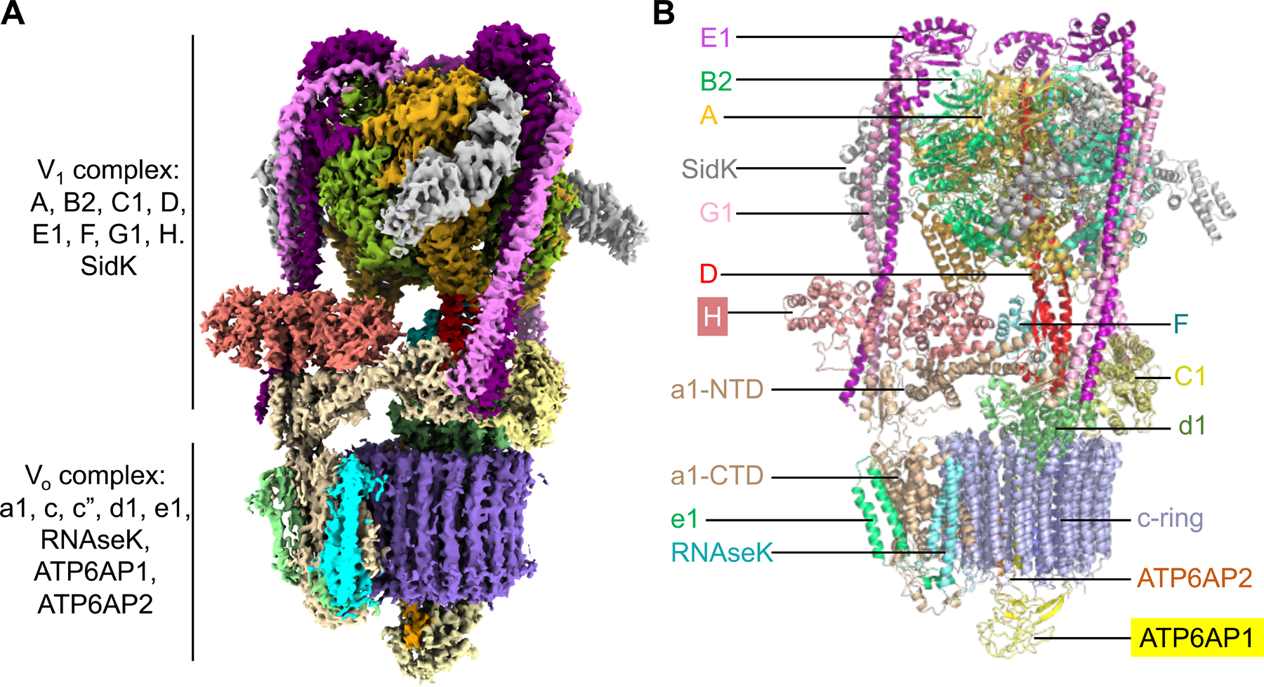
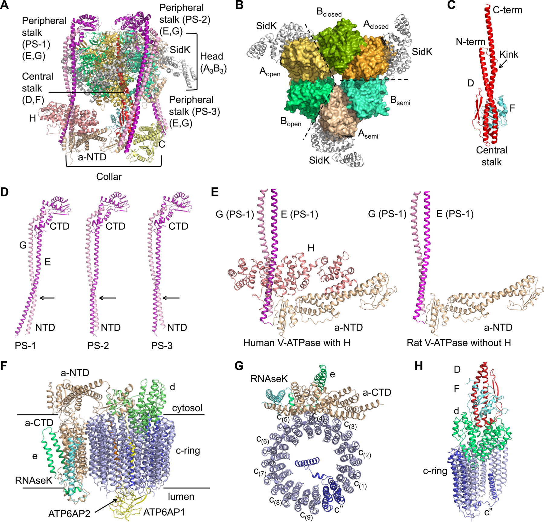
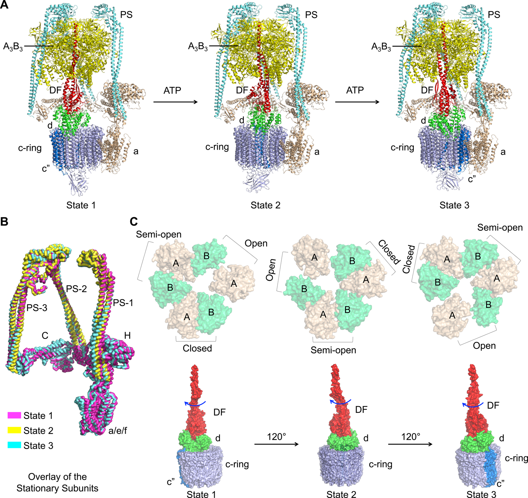
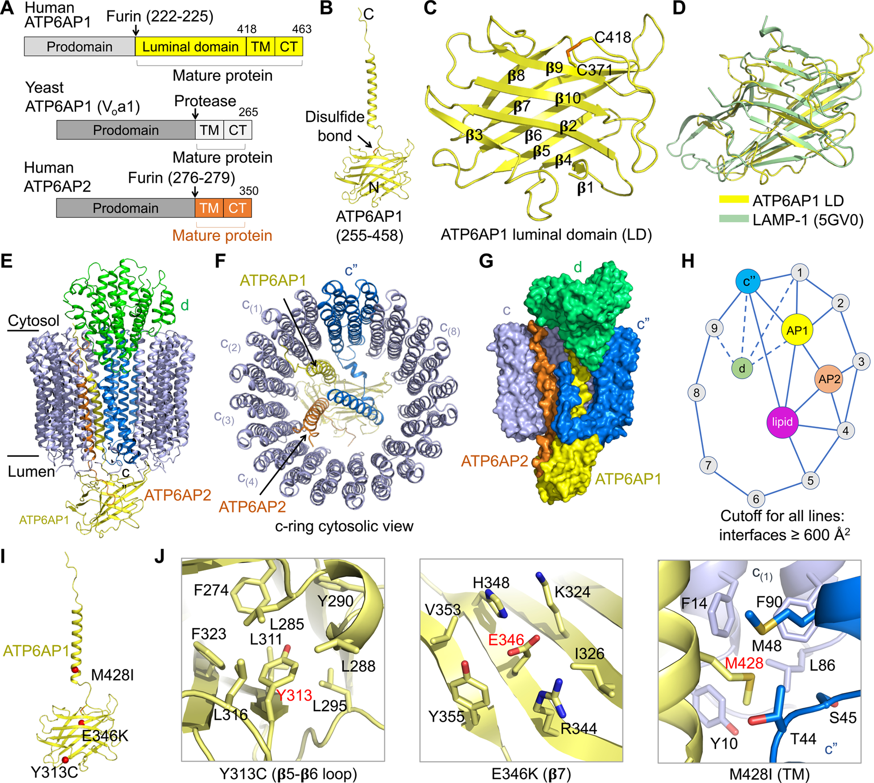
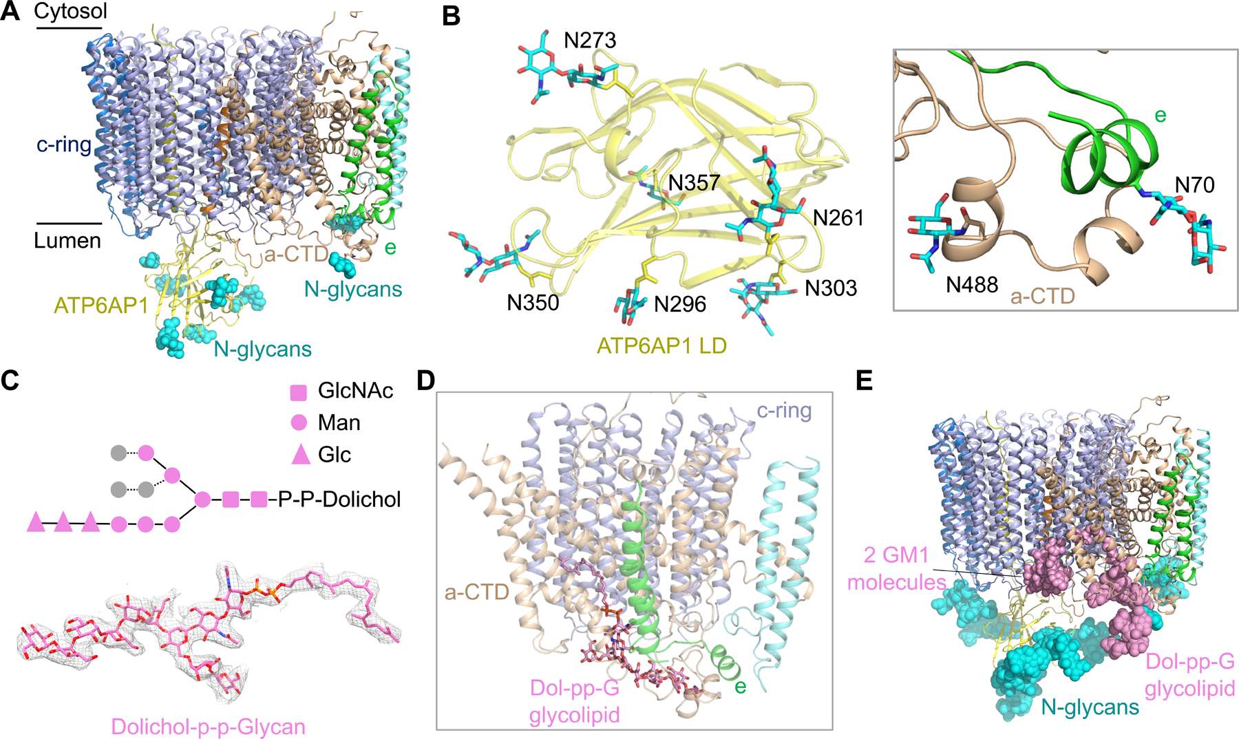
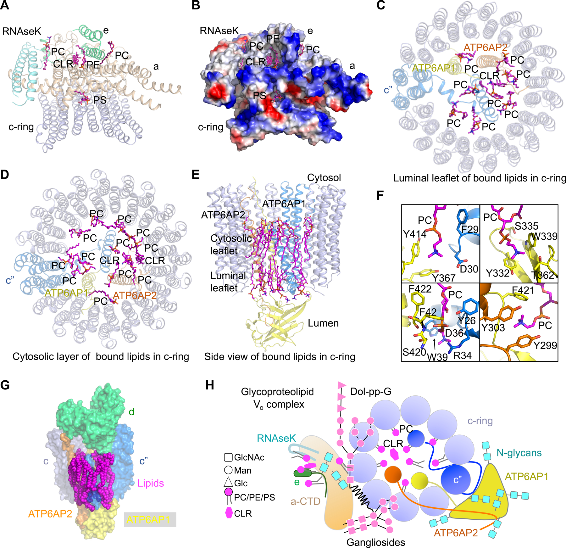
Vesicular- or vacuolar-type adenosine triphosphatases (V-ATPases) are ATP-driven proton pumps comprised of a cytoplasmic V1 complex for ATP hydrolysis and a membrane-embedded Vo complex for proton transfer. They play important roles in acidification of intracellular vesicles, organelles, and the extracellular milieu in eukaryotes. Here, we report cryoelectron microscopy structures of human V-ATPase in three rotational states at up to 2.9-Å resolution. Aided by mass spectrometry, we build all known protein subunits with associated N-linked glycans and identify glycolipids and phospholipids in the Vo complex. We define ATP6AP1 as a structural hub for Vo complex assembly because it connects to multiple Vo subunits and phospholipids in the c-ring. The glycolipids and the glycosylated Vo subunits form a luminal glycan coat critical for V-ATPase folding, localization, and stability. This study identifies mechanisms of V-ATPase assembly and biogenesis that rely on the integrated roles of ATP6AP1, glycans, and lipids.


Conflict of interest statement

Declaration of Interests H.W. is a member of the Scientific Advisory Board of Cell. The other authors declare no competing interests.

Figures

Figure 1.
Figure 1.. Cryo-EM Structure of Human V-ATPase
(A) Cryo-EM density of human V-ATPase (state 1) with subunits color coded. (B) Ribbon diagram of human V-ATPase structure (state 1) with subunits color coded and labeled. Subunits H and ATP6AP1 which are absent or not fully traced in the rat V-ATPase structure (15) are highlighted. See also Table S1 and Figures S1 and S2.
Figure 2.
Figure 2.. Assembly of the V1 and Vo Complex
(A) Ribbon diagram of the V1 complex with each subunit colored as in Figure 1. (B) The A3B3 hexamer surrounded by SidK (grey). The A3B3 hexamer is composed of three pairs of AB heterodimer in three conformations, closed, semi-open (semi) and open. (C) Ribbon diagram of the central stalk that is composed of subunit D (red) and F (cyan). The proline containing kink in subunit D is indicated by an arrow. (D) Conformational differences of the three peripheral stalks, with the CTDs of subunit E (purple) and subunit G (pink) aligned. (E) Comparison between human and rat V-ATPase structures at the region where H is localized. (F) Ribbon diagram of the Vo complex with each subunit colored as in Figure 1. (G) Ribbon diagram of c-ring (blue) with CTD of subunit a (wheat), e (green) and RNAseK (cyan). The c” subunit is colored in marine. (H) Interactions of subunit d (green) with the DF stalk (red and cyan) and the c-ring (blue). See also Figure S3.
Figure 3.
Figure 3.. Molecular Coupling between V1 and Vo Complexes
(A) Ribbon diagrams of the three rotational states of human V-ATPase with the central stalks in red, peripheral stalks in cyan, A3B3 head in tv-yellow, C, H, a, e and RNAseK in wheat, c-ring, ATP6AP1 and ATP6AP2 in blue, and d in green. (B) Overlaid stationary subunits of human V-ATPase structures in three rotational states (state 1 in magenta, state 2 in yellow, and state 3 in cyan). (C) Coordinated changes from the A3B3 head to the rotary DF-d-c-ring unit, shown in surface representations. The CTD hexamers of the A3B3 head (in wheat and green, top) are viewed from the Vo side, displaying the conformational precession between the states. The rotary units containing the central stalk (red), d (green) and the c-ring (blue) (bottom) are presented as side views, showing the ~120° rotation between states in response to the conformational precession at the A3B3 head.
Figure 4.
Figure 4.. Structure of ATP6AP1 and Its Interaction Network
(A) Domain diagrams of human ATP6AP1 and ATP6AP2, and yeast ATP6AP1. The cleavage sites for furin or another protease in their luminal domains are shown. (B) Ribbon diagram of mature ATP6AP1 in the human V-ATPase structure. (C) Ribbon diagram of the luminal domain of ATP6AP1. Location of a conserved disulfide bond between C371 and C418 is marked. Secondary structures in the β-prism fold are labeled. (D) Structural alignment of ATP6AP1 luminal domain (LD, yellow) with LAMP-1 (green) obtained from the DALI server (Holm and Sander, 1995). (E, F) Side view (E) and cytosolic view (F) of the c-ring (c” in blue and c subunits in slate), ATP6AP1 (yellow), ATP6AP2 (orange), and subunit d (green), showing the overall interactions of ATP6AP1 with neighboring subunits. (G) Surface representation of the c-ring, ATP6AP1, ATP6AP2, and subunit d, showing their interactions. (H) A schematic diagram of the interaction network involving c-ring subunits, ATP6AP1, ATP6AP2, subunit d, and lipids. An interaction has to bury > 600 Å2 surface area to qualify. ATP6AP1 is the most connected subunit in the diagram. (I) Missense disease mutations of ATP6AP1 (red) mapped onto its structure. (J) Detailed interactions of the mutation sites within the ATP6AP1 subunit (Y313 and E346) and with neighboring subunits. See also Figure S4.
Figure 5.
Figure 5.. N-linked Glycosylation and Glycolipids of the Vo Complex
(A) N-linked glycans (cyan spheres) at the luminal side of the Vo complex from subunits ATP6AP1, a-CTD and e. (B) Detailed structures of N-linked glycan units on subunits ATP6AP1, a-CTD and e. (C) Diagram of the chemical structure, the model (in stick) and the cryo-EM density (2.0 σ) of the glycolipid dolichol-P-P-glycan (Dol-pp-G). Two copies of N-acetylglucosamine (GlcNAc) (square), six mannose (circle), and three glucose (triangle) units were resolved in the cryo-EM density map. (D) Dol-pp-G bound to subunits c, a-CTD and e. (E) The Vo complex shown with observed glycans at the luminal side. For each N-linked glycosylation site, a total of nine sugar units were modeled based on the complex-type N-glycan structure. GM1: monosialoganglioside. See also Figure S5.
Figure 6.
Figure 6.. Lipid Molecules in the Vo Complex
(A) Lipid molecules at the interfaces of subunits a, e, RNAseK, and the c-ring. These lipids establish extensive interactions with the protein subunits above. PC: phosphatidylcholine; PE: phosphatidylethanolamine; PS: phosphatidylserine; CLR: cholesterol. (B) Electrostatic surface representation of subunits a, e, RNAseK, and c-ring, showing the interactions between the lipids and protein subunits. (C, D) Lipid molecules inside the c-ring at the luminal leaflet (C) and the cytosolic leaflet (D). These lipids establish extensive interactions with ATP6AP1, ATP6AP2, and the c-ring. (E) Side view of bound lipids inside the c-ring. (F) Detailed interactions of lipid molecules with ATP6AP1 (yellow), c” (blue) and ATP6AP2 (orange). (G) Surface representation of lipid molecules, c-ring, ATP6AP1, ATP6AP2, and subunit d, showing their interactions. (H) A schematic diagram of the luminal view of the glycoproteolipid Vo complex, showing all protein subunits, N-linked glycans, glycolipids, and ordered PC and cholesterol lipids inside and outside the c-ring. See also Figure S6.

Comment in

Similar articles

Cited by

References

    1. Abbas YM, Wu D, Bueler SA, Robinson CV, and Rubinstein JL (2020). Structure of V-ATPase from the mammalian brain. Science 367, 1240–1246. - PMC - PubMed
    1. Adams PD, Afonine PV, Bunkoczi G, Chen VB, Davis IW, Echols N, Headd JJ, Hung LW, Kapral GJ, Grosse-Kunstleve RW, et al. (2010). PHENIX: a comprehensive Python-based system for macromolecular structure solution. Acta Crystallogr D Biol Crystallogr 66, 213–221. - PMC - PubMed
    1. Biasini M, Bienert S, Waterhouse A, Arnold K, Studer G, Schmidt T, Kiefer F, Cassarino TG, Bertoni M, Bordoli L, et al. (2014). SWISS-MODEL: modelling protein tertiary and quaternary structure using evolutionary information. Nucleic acids research 42, W252–258. - PMC - PubMed
    1. Bolla JR, Agasid MT, Mehmood S, and Robinson CV (2019). Membrane Protein-Lipid Interactions Probed Using Mass Spectrometry. Annu Rev Biochem 88, 85–111. - PubMed
    1. Casey JR, Grinstein S, and Orlowski J (2010). Sensors and regulators of intracellular pH. Nat Rev Mol Cell Biol 11, 50–61. - PubMed

Publication types

MeSH terms

Substances