. 2020 Dec 21;30(24):5049-5057.e3.
doi: 10.1016/j.cub.2020.09.061. Epub 2020 Oct 15.

An Axon-Pathfinding Mechanism Preserves Epithelial Tissue Integrity

Affiliations

An Axon-Pathfinding Mechanism Preserves Epithelial Tissue Integrity

Christian Cammarota et al. Curr Biol. .

Abstract

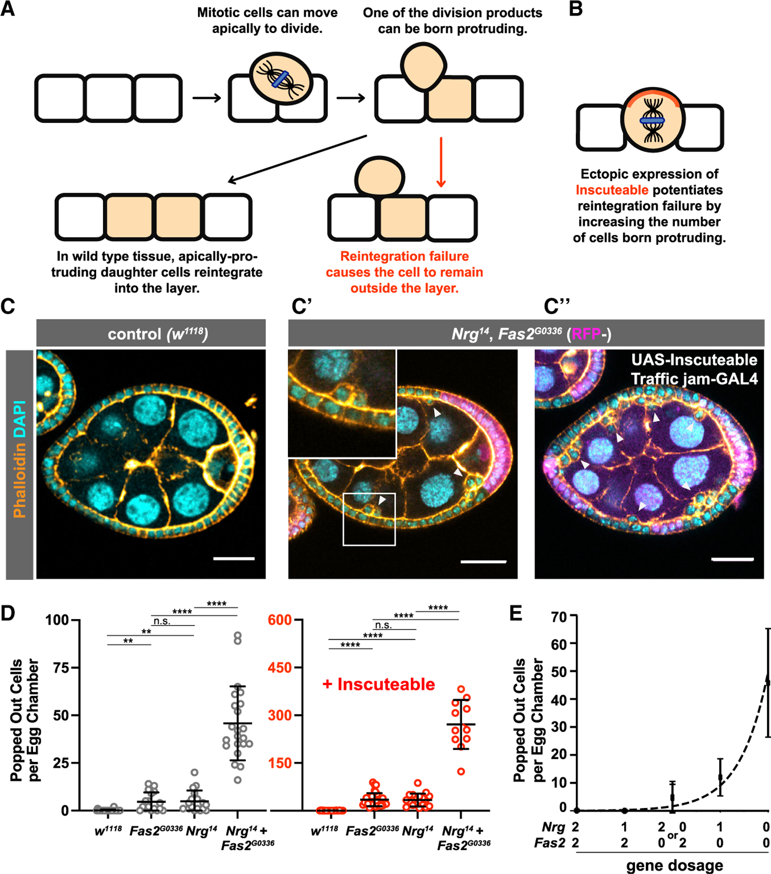
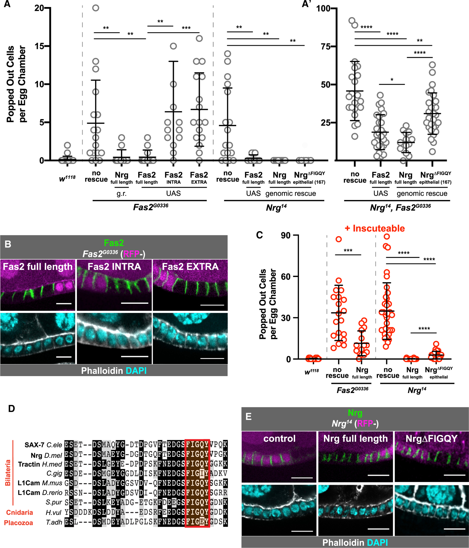
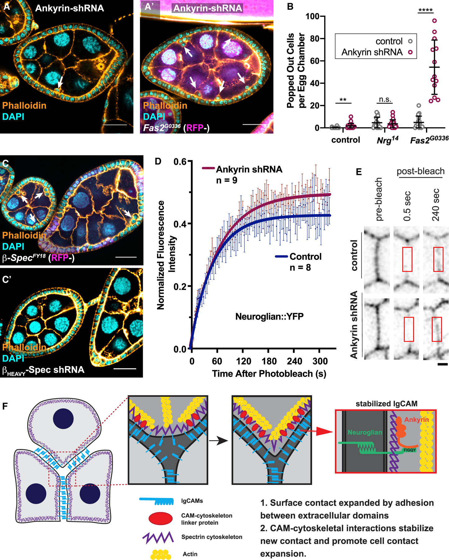
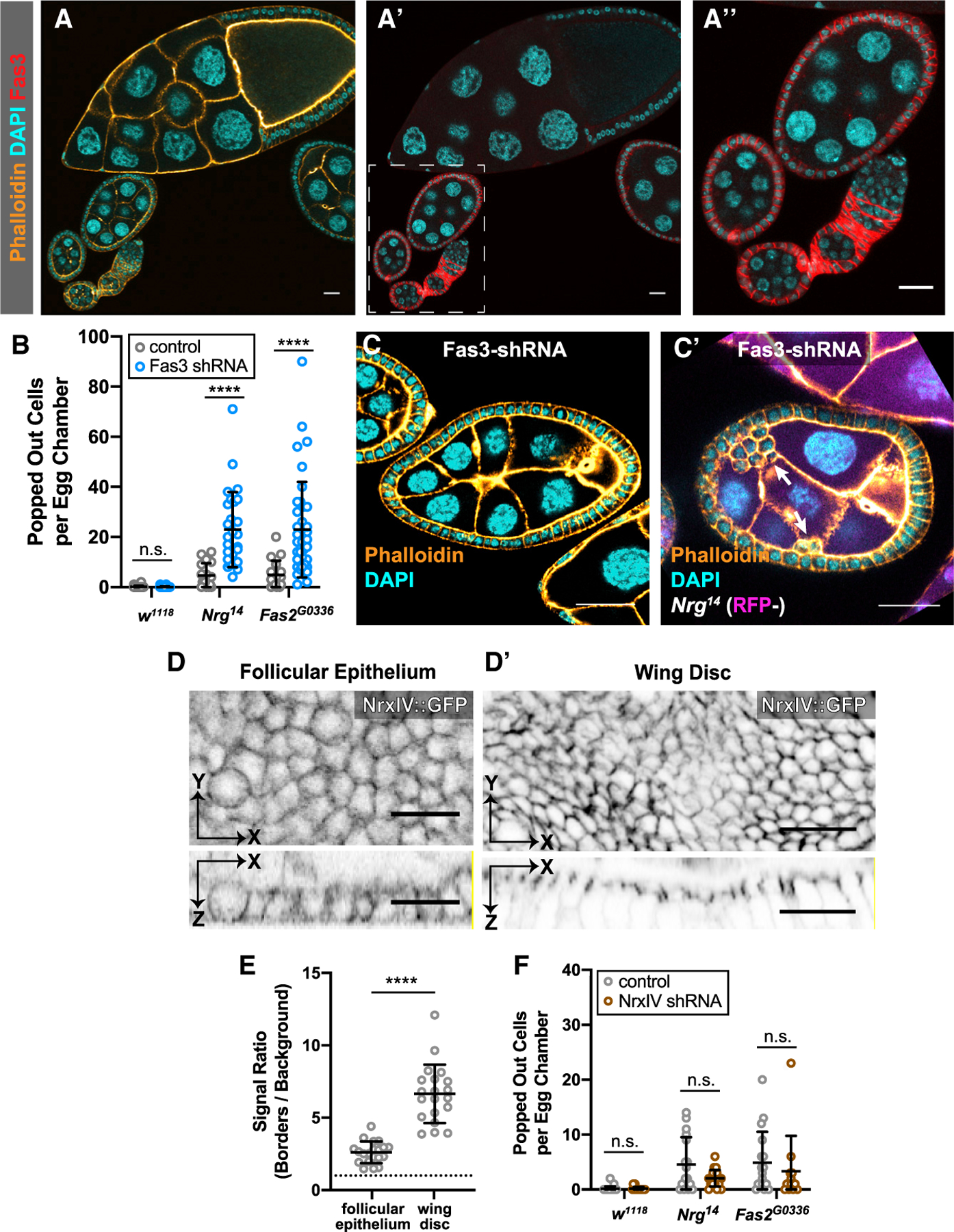
Epithelial tissues form the boundaries of organs, where they perform a range of functions, including secretion, absorption, and protection. These tissues are commonly composed of discrete cell layers-sheets of cells that are one-cell thick. In multiple systems examined, epithelial cells round up and move in the apical direction before dividing, likely in response to neighbor-cell crowding [1-6]. Because of this movement, daughter cells may be born displaced from the tissue layer. Reintegration of these displaced cells supports tissue growth and maintains tissue architecture [4]. Two conserved IgCAMs (immunoglobulin superfamily cell adhesion molecules), neuroglian (Nrg) and fasciclin 2 (Fas2), participate in cell reintegration in the Drosophila follicular epithelium [4]. Like their vertebrate orthologs L1CAM and NCAM1/2, respectively, Nrg and Fas2 are cell adhesion molecules primarily studied in the context of nervous system development [7-10]. Consistent with this, we identify another neural IgCAM, Fasciclin 3 (Fas3), as a reintegration factor. Nrg, Fas2, and Fas3 are components of the insect septate junction, the functional equivalent of the vertebrate tight junction, but proliferating follicle cells do not have mature septate junctions, and we find that the septate junction protein neurexin IV does not participate in reintegration [11, 12]. Here, we show that epithelial reintegration works in the same way as IgCAM-mediated axon growth and pathfinding; it relies not only on extracellular adhesion but also mechanical coupling between IgCAMs and the lateral spectrin-based membrane skeleton. Our work indicates that reintegration is mediated by a distinct epithelial adhesion assembly that is compositionally and functionally equivalent to junctions made between axons.

Keywords: adhesion; epithelia; epithelial junctions; reintegration.


Conflict of interest statement

Declaration of Interests The authors declare no competing interests.

Figures

Figure 1.
Figure 1.. Nrg and Fas2 Act in Parallel to Drive Reintegration
(A) Diagram showing apical cell movement during mitosis and subsequent cell reintegration. (B) Inscuteable increases out-of-plane divisions and therefore the number of reintegration events. (C and D) Genetic disruption of both Fas2 and Nrg causes a synergistic increase in reintegration failure over disruption of either gene alone. Ectopic expression of Inscuteable has a multiplicative effect on reintegration failure. Representative images are shown in (C) and quantification in (D). Each data point represents the number of extralayer (popped-out) cells in the entirety of one egg chamber. Each column represents three or more dissections of five or more flies. Significance was determined using an unpaired, two-tailed Student’s t test with Welch’s correction. p values left to right: p = 0.0018, p = 0.0031, p = 0.8723, p < 0.0001, p < 0.0001, p < 0.0001, p < 0.0001, p = 0.8201, p < 0.0001, p < 0.0001. Average and error bars correspond to mean and SD. Scale bars in (C) correspond to 20 µm. (E) The relationship between reintegration failure and gene function is not linear. A plot of gene dosage versus misplaced cell number fits an exponential curve (R2 = 0.972), providing a speculative model for reintegration function. Average and error bars correspond to mean and SD. See also Video S1 and Figure S1.
Figure 2.
Figure 2.. Dissection of Nrg and Fas2 Function
(A) Genetic rescue experiments reveal the relative roles of Nrg and Fas2 in reintegration, through assessment of the “popping out” phenotype. Average and error bars correspond to mean and SD. Expression of full-length Fas2 rescues reintegration failure in Nrg- and Fas2-null tissue, but both the extracellular and intracellular domains are required to rescue Fas2G0336. Neuroglian rescues reintegration failure in Nrg- and Fas2-null tissue. Removal of the FIGQY subsequence from the epithelial Nrg isoform allows for partial rescue in Nrg-null tissue. p values left to right: p = 0.0052, p = 0.0052, p = 0.0071, p = 0.0001, p = 0.0023, p = 0.0014, p = 0.0014. (A’) Neither expression of full-length Fas2 or Nrg fully rescues reintegration failure caused by Nrg14,Fas2G0336 double mutants. The Nrg variant with a disruption in the epithelial FIGQY subsequence rescues reintegration failure significantly less than the full Nrg rescue. UAS-Fas2-YFP and variants were expressed using the driver Traffic Jam-GAL4. Neuroglian and Nrg variants were expressed from a single genomic rescue construct. p values left to right: p < 0.0001, p < 0.0001, p = 0.0044, p = 0.0221, p < 0.0001. (B) Fas2 localizes to cell-cell borders without either its extracellular or intracellular domain. UAS-Fas2-YFP and its truncated variants, expressed using Traffic Jam-Gal4, could all be detected at follicle cell-cell borders, whether in the presence (RFP+) or absence (RFP−) of endogenous Fas2. (C) Ectopic expression of Insc reveals that both Nrg overexpression in Fas2-null and expression of Nrg missing the FIGQY subsequence in Nrg-null tissues incompletely rescue the reintegration failure phenotype. Average and error bars correspond to mean and SD. p values left to right: p = 0.0002, p < 0.0001, p < 0.0001, p < 0.0001. (D) The ankyrin-binding domain of Nrg/L1CAM is evolutionarily conserved. (E) The FIGQY subsequence is not required for Nrg localization to epithelial cell-cell borders. Representative images are shown in (E) and quantified in Figure S2C. Quantification and statistical tests in (A) and (C) were performed as in Figure 1. Average and error bars in (A) and (C) correspond to mean and SD. Scale bars in (B) and (E) correspond to 5 µm. See also Figure S2.
Figure 3.
Figure 3.. Nrg and Ankyrin Cooperate to Drive Cell Reintegration
(A and B) Genetic disruption of ankyrin results in failed cell reintegration. Expression of ankyrin shRNA, driven by Traffic Jam-GAL4, results in ~2 popped-out cells per egg chamber. Ankyrin shRNA potentiates the reintegration failure phenotype seen in Fas2-null tissue, but not Nrg-null tissue. Representative images are shown in (A) and quantification in (B). Quantification and statistical tests were performed as in Figure 1. Scale bars in (A) correspond to 20 µm. Average and error bars correspond to mean and SD. p values from left to right: p = 0.0086, p = 0.4663, p < 0.0001. (C) Popped-out cells, indicating reintegration failure, are apparent in FE tissue mutant for β-spectrinFY18, but not βHEAVY-spectrin. Scale bars correspond to 20 µm. (D) Fluorescence recovery curves of Nrg::YFP after photobleaching. Lateral FE cell junctions were bleached in egg chambers expressing ankyrin-shRNA driven by Traffic Jam-GAL4 or egg chambers with the driver alone (control). Fluorescence intensity was normalized to account for inherent photobleaching due to imaging. Experiments were performed independently with number of experimental repeats indicated. Curves were calculated by fitting a one-phase association curve. Error bars represent SEM. The 95% confidence interval (CI) of the control plateau is 41–44. This does not overlap with the 95% CI of the ankyrin-shRNA plateau, which is 48–51. (E) Frames from an example FRAP experiment, showing the fluorescence of Nrg::YFP at a cell junction prior to and post bleaching in control and ankyrin-shRNA-expressing egg chambers. Red boxes indicate photobleached region over which fluorescence intensity was quantified. Scale bars correspond to 1 µm. (F) We propose a refined model for IgCAM-mediated cell reintegration in epithelia. Both extracellular adhesion and intracellular connection to the cytoskeleton is necessary for the reintegration of cells into epithelial layers. Specifically, we reveal that the spectrin-based cytoskeleton stabilizes Nrg cell-cell adhesion through ankyrin to facilitate reintegration. We propose that the SBMS mechanically stabilizes the trans-interactions of Nrg at the “leading edge” of integrating cells to facilitate the progression of cell reintegration. Nrg-ankyrin interactions thereby provide a traction force (grip). See also Figure S3.
Figure 4.
Figure 4.. Fas3 Participates in Reintegration, but NrxIV does Not
(A) Fas3 localizes to follicle cell-cell borders in egg chambers prior to stage 7. Fas3 immunoreactivity is detected in early egg chambers, in which the follicular epithelium is proliferative (stages 1–6, in dashed box), but drops off substantially by mid-stage (stage 10, top of panel). (B and C) Fas3 disruption potentiates reintegration failure in Nrg and Fas2 mutant tissue. Fas3 knockdown does not result in misplaced cells (B) unless combined with mutation of either Nrg or Fas2 (B and C). Fas3-shRNA was driven with Traffic Jam-GAL4. Misplaced cells were quantified as in Figure 1. Average and error bars correspond to mean and SD. p values left to right: p = 0.5948, p < 0.0001, p < 0.0001. (D and E) NrxIV is weakly localized in the early follicular epithelium. NrxIV::GFP, a protein trap, demonstrates weaker junctional localization in the follicular epithelium (D) than in the mature wing disc (D’) relative to local background signal (quantification in E). Signal intensity was measured at septate junctions (wing disc) or along lateral cell-cell contacts (follicular epithelium) and compared with adjacent cytoplasmic signal. Average and error bars represent mean and SD. Significance was determined using an unpaired, two-tailed Student’s t test with Welch’s correction p < 0.0001. (F) NrxIV disruption does not enhance reintegration failure. NrxIV-shRNA, driven with Traffic Jam-GAL4, does not increase the number of misplaced cells in control tissue or in Nrg or Fas2 mutant tissue. Average and error bars represent mean and SD. p values from left to right: p = 0.9166, p = 0.0547, p = 0.5095. Quantification and statistical analysis were performed as in Figure 1. All scale bars correspond to 20 µm. See also Figure S4.

Comment in

  • Cell Biology: Pardon the Intrusion.
    Williams SE, Lough KJ. Williams SE, et al. Curr Biol. 2020 Dec 21;30(24):R1481-R1484. doi: 10.1016/j.cub.2020.10.036. Curr Biol. 2020. PMID: 33352130

Similar articles

Cited by

References

    1. Sauer FC (1936). The interkinetic migration of embryonic epithelial nuclei. J. Morphol 60, 1–11.
    1. Spear PC, and Erickson CA (2012). Interkinetic nuclear migration: a mysterious process in search of a function. Dev. Growth Differ 54, 306–316. - PMC - PubMed
    1. Packard A, Georgas K, Michos O, Riccio P, Cebrian C, Combes AN, Ju A, Ferrer-Vaquer A, Hadjantonakis A-K, Zong H, et al. (2013). Luminal mitosis drives epithelial cell dispersal within the branching ureteric bud. Dev. Cell 27, 319–330. - PMC - PubMed
    1. Bergstralh DT, Lovegrove HE, and St Johnston D (2015). Lateral adhesion drives reintegration of misplaced cells into epithelial monolayers. Nat. Cell Biol 17, 1497–1503. - PMC - PubMed
    1. McKinley KL, Stuurman N, Royer LA, Schartner C, Castillo-Azofeifa D, Delling M, Klein OD, and Vale RD (2018). Cellular aspect ratio and cell division mechanics underlie the patterning of cell progeny in diverse mammalian epithelia. eLife 7, 19. - PMC - PubMed

Publication types

MeSH terms