. 2021 Jan 11;31(1):13-24.e4.
doi: 10.1016/j.cub.2020.09.071. Epub 2020 Oct 15.

Mutations in Metabotropic Glutamate Receptor 1 Contribute to Natural Short Sleep Trait

Affiliations

Mutations in Metabotropic Glutamate Receptor 1 Contribute to Natural Short Sleep Trait

Guangsen Shi et al. Curr Biol. .

Abstract

Sufficient and efficient sleep is crucial for our health. Natural short sleepers can sleep significantly shorter than the average population without a desire for more sleep and without any obvious negative health consequences. In searching for genetic variants underlying the short sleep trait, we found two different mutations in the same gene (metabotropic glutamate receptor 1) from two independent natural short sleep families. In vitro, both of the mutations exhibited loss of function in receptor-mediated signaling. In vivo, the mice carrying the individual mutations both demonstrated short sleep behavior. In brain slices, both of the mutations changed the electrical properties and increased excitatory synaptic transmission. These results highlight the important role of metabotropic glutamate receptor 1 in modulating sleep duration.

Keywords: loss-of-function; mGluR1; short-sleep.


Conflict of interest statement

Declaration of Interests The authors declare no competing interests.

Figures

Figure 1.
Figure 1.. Two Mutations Were Identified in Two Unrelated FNSS Families
(A) Pedigree of families (K50230 and K07331) carrying the GRM1 mutations. Circles and squares indicate female and male subjects, respectively. One deceased individual is listed as “probably affected” based on historical report of his spouse and descendants. See also Table S1 for detailed sleep schedules. (B) Daily sleep time distribution of the carriers and non-carriers from the two families. (C) Representative sleep architecture of two FNSS subjects based on the two baseline nights of polysomnography (PSG). Total sleep times measured by PSG during these two nights are shown on the right. (D) Locations of the mutated amino acid residues in the full-length mGluR1. (E) Alignment of mutant residues among vertebrate and mammalian species. **p < 0.01. Two-tailed unpaired Student’s t test. Error bars represent ± SEM.
Figure 2.
Figure 2.. Mutant mGluR1s Show Less Responsiveness to DHPG Based on ERK Activation
(A and B) Representative p-ERK immunoblots for HEK293 cells transfected with WT or mutant mGluR1b (A) and mGluR1a (B). These cells were treated with the indicated doses of DHPG for 5 min before harvesting. See Figure S1 for vector transfected controls. (C and D) Quantified results for (A) and (B). Different colors indicate four independent experiments. *p < 0.05, n.s. = not significant. RM (repeated measures) one-way ANOVA, post hoc Dunnett’s multiple comparisons test (C). Two-tailed paired Student’s t test (D). Error bars represent ± SEM.
Figure 3.
Figure 3.. Mutant mGluR1s Are Less Active in Tango Assays
(A and B) Relative activity of mGluR1a WT-Tango and mGluR1a S458A-Tango in response to DHPG, MCPG (A), and nitazoxanide (B). See also Figure S2B for expression control. (C–E) Relative activity of mGluR1b WT-Tango, mGluR1b R889W-Tango, and mGluR1b S458A-Tango in response to DHPG (C), MCPG (D), and nitazoxanide (E). See also Figure S2C for expression control. (F and G) Relative activity of equal mixture (1:1) expression of mGluR1b WT-Tango and mGluR1b S458A-Tango (F) and mGluR1b WT-Tango and mGluR1b R889W-Tango (G) in response to DHPG. (H) Relative activity of equal mixture (1:1) expression of mGluR1b-Tango and non-Tango constructs in response to DHPG. See Figure S2D for schematic setup. *p < 0.05, ***p < 0.001, ****p < 0.0001. Two-way ANOVA, post hoc Sidak’s multiple comparisons test. Error bars represent ± SEM.
Figure 4.
Figure 4.. Grm1 Mutations Reduce the Total Sleep Time in FNSS Mouse Models
(A–C) Total (A), NREM (B), and REM (C) sleep times by EEG/EMG within 24 h for Grm1 +/+ (n = 16) and Grm1b R889W +/m (n = 11) mice. (D–F) Total (D), NREM (E), and REM (F) sleep times by EEG/EMG within 24 h for Grm1 +/+ (n = 19) and Grm1 S458A +/m (n = 23) mice. See also Figures S3 and S4 for additional analysis. *p < 0.05, n.s. = not significant. Two-tailed Student’s t test. Error bars represent ± SEM.
Figure 5.
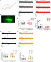
Figure 5.. Grm1 Mutations Change the Electrophysiological Properties of DG Grm1+ Neurons
(A) Schematic of patch-clamp of DG neurons. (B) Representative GFP+ neurons in an acute slice for recording. (C and D) Representative voltage-clamped mEPSC traces before, during, and after DHPG (25 μM) treatment for Grm1 +/+ (C) and Grm1b R889W +/m (D) neurons. (E and F) Summary of mEPSC amplitude (E) and frequency (F) before, during, and after DHPG (25 μM) treatment for Grm1 +/+ (n = 14 cells) and Grm1b R889W +/m (n = 12 cells) neurons. (G–J) The same as (C–F) except Grm1 S458A +/m is shown instead of Grm1b R889W. Grm1 +/+ (n = 12 cells) and Grm1 S458A +/m (n = 15 cells). See Figure S5 for GFP positive cells in other brain regions. See also Figure S6 and Tables S2, S3, and S4 for additional electrophysiology analysis. *p < 0.05, **p < 0.01, n.s. = not significant. Two-tailed paired Student’s t test.
Figure 6.
Figure 6.. Grm1 Mutations Change the Transcriptome in the Hippocampus
(A) Heatmap of genes with >1.5-fold changes between WT (n = 4, left 4 columns) and Grm1b R889W +/m hippocampus (n = 4, middle four columns). The same genes from Grm1 S458A +/m tissues (n = 4) were aligned in the right four columns. Blue and red lines at the left side indicate downregulated and upregulated gene clusters respectively. (B) Heatmap of genes with >1.5-fold changes between WT (n = 4, left four columns) and Grm1 S458A+/m hippocampus (n = 4, middle four columns). The same genes from Grm1b R889W +/m tissues (n = 4) were aligned in the right four columns. (C–F) Summary of genes that showed the same or similar (trend) increase (C and D) or decrease (E and F) between Grm1b R889W +/m (RW) and Grm1 S458A +/m (SA) tissues. See Data S1 for the names, fold changes, and p values of genes in (C–F).

Similar articles

Cited by

References

    1. Hirshkowitz M, Whiton K, Albert SM, Alessi C, Bruni O, DonCarlos L, Hazen N, Herman PJ, Adams Hillard PJ, Katz ES, et al. (2015). National Sleep Foundation’s updated sleep duration recommendations: final report. Sleep Health 1, 233–243. - PubMed
    1. He Y, Jones CR, Fujiki N, Xu Y, Guo B, Holder JL Jr., Rossner MJ, Nishino S, and Fu YH (2009). The transcriptional repressor DEC2 regulates sleep length in mammals. Science 325, 866–870. - PMC - PubMed
    1. Ashbrook LH, Krystal AD, Fu YH, and Ptáček LJ (2020). Genetics of the human circadian clock and sleep homeostat. Neuropsychopharmacology 45, 45–54. - PMC - PubMed
    1. Shi G, Xing L, Wu D, Bhattacharyya BJ, Jones CR, McMahon T, Chong SYC, Chen JA, Coppola G, Geschwind D, et al. (2019). A Rare Mutation of β1-Adrenergic Receptor Affects Sleep/Wake Behaviors. Neuron 103, 1044–1055.e7. - PMC - PubMed
    1. Xing L, Shi G, Mostovoy Y, Gentry NW, Fan Z, McMahon TB, Kwok PY, Jones CR, Ptáček LJ, and Fu YH (2019). Mutant neuropeptide S receptor reduces sleep duration with preserved memory consolidation. Sci. Transl. Med. 11, eaax2014. - PMC - PubMed

Publication types

Substances