

Multi-task deep learning based CT imaging analysis for COVID-19 pneumonia: Classification and segmentation
- PMID: 33065387
- PMCID: PMC7543793
- DOI: 10.1016/j.compbiomed.2020.104037
Multi-task deep learning based CT imaging analysis for COVID-19 pneumonia: Classification and segmentation
Abstract
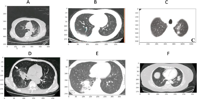
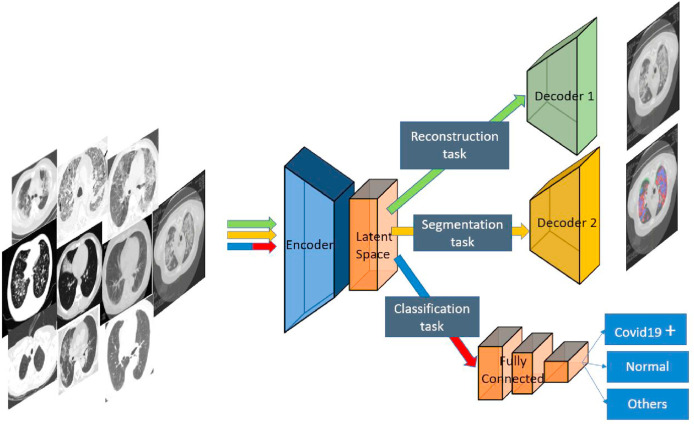
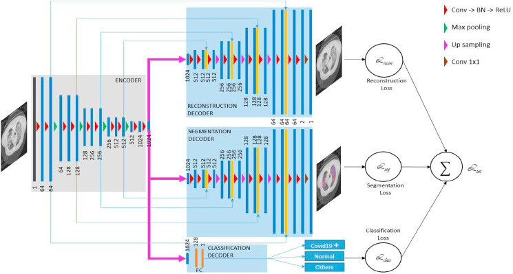
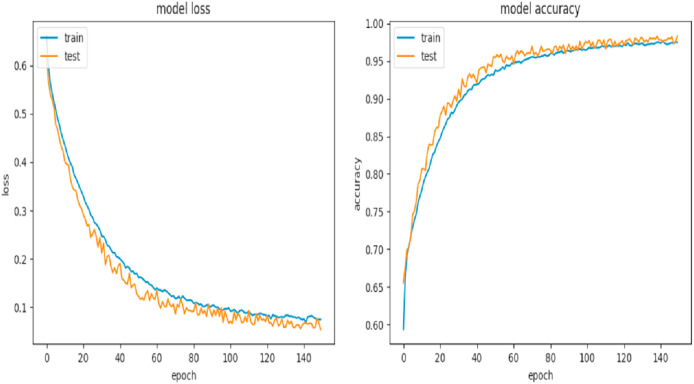
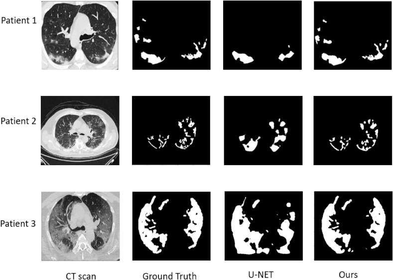
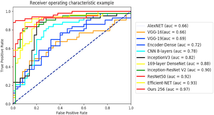
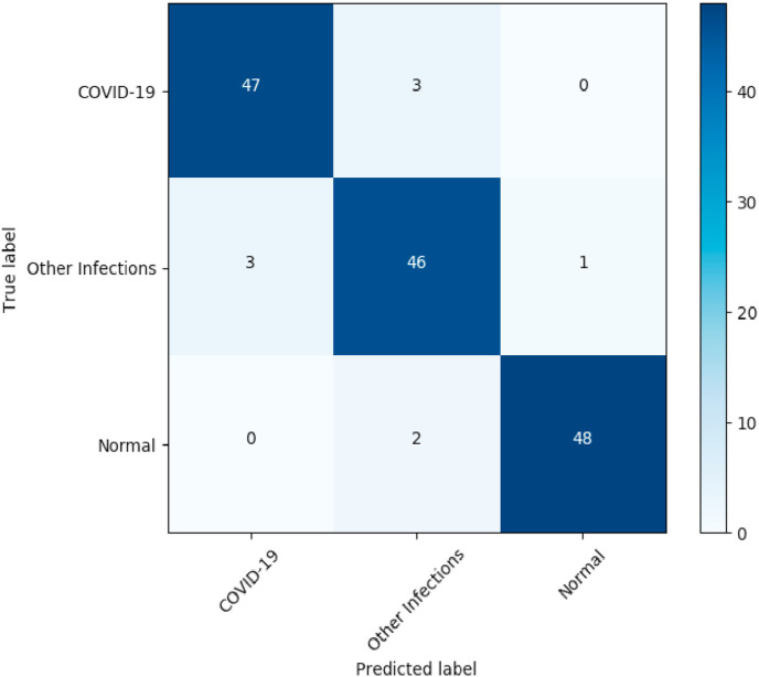
This paper presents an automatic classification segmentation tool for helping screening COVID-19 pneumonia using chest CT imaging. The segmented lesions can help to assess the severity of pneumonia and follow-up the patients. In this work, we propose a new multitask deep learning model to jointly identify COVID-19 patient and segment COVID-19 lesion from chest CT images. Three learning tasks: segmentation, classification and reconstruction are jointly performed with different datasets. Our motivation is on the one hand to leverage useful information contained in multiple related tasks to improve both segmentation and classification performances, and on the other hand to deal with the problems of small data because each task can have a relatively small dataset. Our architecture is composed of a common encoder for disentangled feature representation with three tasks, and two decoders and a multi-layer perceptron for reconstruction, segmentation and classification respectively. The proposed model is evaluated and compared with other image segmentation techniques using a dataset of 1369 patients including 449 patients with COVID-19, 425 normal ones, 98 with lung cancer and 397 of different kinds of pathology. The obtained results show very encouraging performance of our method with a dice coefficient higher than 0.88 for the segmentation and an area under the ROC curve higher than 97% for the classification.
Keywords: Computed tomography images; Coronavirus (COVID-19); Deep learning; Image classification; Image segmentation; Multitask learning.
Copyright © 2020 Elsevier Ltd. All rights reserved.
Conflict of interest statement
None Declared.
Figures


References
-
- Abbasian Ardakani A., Bitarafan-Rajabi A., Mohammadzadeh A., Mohammadi A., Riazi R., Abolghasemi J., Homayoun Jafari A., Bagher Shiran M. A hybrid multilayer filtering approach for thyroid nodule segmentation on ultrasound images. J. Ultrasound Med. 2019;38:629–640. - PubMed
-
- Amyar A., Ruan S., Gardin I., Chatelain C., Decazes P., Modzelewski R. 3-d rpet-net: development of a 3-d pet imaging convolutional neural network for radiomics analysis and outcome prediction. IEEE Transactions on Radiation and Plasma Medical Sciences. 2019;3:225–231.
-
- Amyar A., Ruan S., Gardin I., Herault R., Clement C., Decazes P., Modzelewski R. Radiomics-net: convolutional neural networks on fdg pet images for predicting cancer treatment response. J. Nucl. Med. 2018;59 324–324.
-
- Badrinarayanan V., Kendall A., Cipolla R. Segnet: a deep convolutional encoder-decoder architecture for image segmentation. IEEE Trans. Pattern Anal. Mach. Intell. 2017;39:2481–2495. - PubMed
MeSH terms
LinkOut - more resources
Full Text Sources
Other Literature Sources
Medical
