. 2021 Feb;51(2):471-482.
doi: 10.1002/eji.201948451. Epub 2020 Nov 16.

Dysregulated RASGRP1 expression through RUNX1 mediated transcription promotes autoimmunity

Affiliations

Dysregulated RASGRP1 expression through RUNX1 mediated transcription promotes autoimmunity

Matthijs J D Baars et al. Eur J Immunol. 2021 Feb.

Abstract

RasGRP1 is a Ras guanine nucleotide exchange factor, and an essential regulator of lymphocyte receptor signaling. In mice, Rasgrp1 deletion results in defective T lymphocyte development. RASGRP1-deficient patients suffer from immune deficiency, and the RASGRP1 gene has been linked to autoimmunity. However, how RasGRP1 levels are regulated, and if RasGRP1 dosage alterations contribute to autoimmunity remains unknown. We demonstrate that diminished Rasgrp1 expression caused defective T lymphocyte selection in C57BL/6 mice, and that the severity of inflammatory disease inversely correlates with Rasgrp1 expression levels. In patients with autoimmunity, active inflammation correlated with decreased RASGRP1 levels in CD4+ T cells. By analyzing H3K27 acetylation profiles in human T cells, we identified a RASGRP1 enhancer that harbors autoimmunity-associated SNPs. CRISPR-Cas9 disruption of this enhancer caused lower RasGRP1 expression, and decreased binding of RUNX1 and CBFB transcription factors. Analyzing patients with autoimmunity, we detected reduced RUNX1 expression in CD4+ T cells. Lastly, we mechanistically link RUNX1 to transcriptional regulation of RASGRP1 to reveal a key circuit regulating RasGRP1 expression, which is vital to prevent inflammatory disease.

Keywords: RASGRP1; RUNX1; T cells; autoimmunity; transcription.


Conflict of interest statement

Jeroen Roose is a co‐founder and scientific advisor of Seal Biosciences, Inc. and on the scientific advisory committee for the Mark Foundation for Cancer research. A.M. is a cofounder, member of the Boards of Directors and a member of the Scientific Advisory Boards of Spotlight Therapeutics and Arsenal Biosciences. A.M. has served as an advisor to Juno Therapeutics, was a member of the scientific advisory board at PACT Pharma, and was an advisor to Trizell. A.M. owns stock in Arsenal Biosciences, Spotlight Therapeutics and PACT Pharma. The Marson lab has received research support from Juno Therapeutics, Epinomics, Sanofi, GlaxoSmithKline, Gilead, and Anthem. D.R.S. is a co‐founder of Beeline Therapeutics. The other authors have no commercial or financial interests.

Figures

Figure 1
Figure 1
Limited RasGRP1 expression results in dose‐dependent autoimmunity in mice. (A) Western blot was performed to analyze RasGRP1, and α‐tubulin (loading control) protein levels expressed by thymocytes of 11 weeks old mice with different genotypes: +/+ (WT, N = 4), +/− (Rasgrp1 heterozygote, N = 3), −/− (Rasgrp1 KO, N = 4), 1 experiment. (B and C) Anti‐nuclear antibody presence (ANA) was determined by Hep2 assays in sera isolated from mice at 11 weeks old (B, N = 12, N = 9, N = 6), and 24 weeks old (C, N = 11, N = 13, N = 10), 2 separate experiments. Bars depict total numbers of mice tested positive (black), and negative (white), percentages of ANA positive samples are indicated. Fisher's exact test was used. * p < 0.05, ** p < 0.01, *** p < 0.001, **** p < 0.0001.
Figure 2
Figure 2
RASGRP1 levels and expression regulation are associated with human autoimmunity. (A) Displayed are qPCR analyses of RASGRP1 mRNA levels, normalized to B2M in CD4+ T cells that were isolated from juvenile idiopathic arthritis patients disease remission blood (JIA‐Remission, N = 8, in three experiments of N = 3 or N = 2), active inflammatory disease blood (JIA‐Active, N = 8, in three experiments of N = 3 or N = 2), or synovial fluid (JIA‐Active synovial fluid, N = 3, one experiment), and healthy adult donors blood (N = 6, in two experiments of N = 3). Shown are averages ± SD. 2–dCT values of RASGRP1 corrected for B2M. One‐way ANOVA, with Holm‐Sidak's multiple comparisons test was used for statistical analysis, * p < 0.05, ** p < 0.01. (B) RASGRP1 locus showing fine mapped disease‐associated SNPs for Crohn's disease (CD) [14], Type 1 diabetes (T1D) [15], and rheumatoid arthritis (RA) [13]. The GWAS data were overlapped with histone 3 lysine 27 acetylation (H3K27Ac) tracks for different T cell types (Roadmap Epigenomics Project [44]): Naïve conventional T cells (Tconv Naïve), anti‐CD3/anti‐CD28 stimulated conventional T cells (Tconv Stim), and CD4+FOXP3+ regulatory T cells (Treg). 2 regions with H3K27 signal and colocalizing autoimmune SNPs are highlighted as autoimmunity‐associated enhancer regions.
Figure 3
Figure 3
Autoimmunity‐associated enhancer 1 regulates RASGRP1 expression. (A) CRISPR‐Cas9 ribonucleoprotein editing of exon 2 was performed in Jurkat cells. Displayed is a western blot showing RasGRP1 and α‐tubulin (loading control), parental Jurkat cells, one non‐edited clone (unedited‐i), and one RASGRP1‐KO clone (RG1‐KO), 3 separate experiments. (B) CRISPR‐Cas9 targeting of autoimmunity‐associated enhancer 1 was performed in Jurkat cells. Displayed are the sequences of two mutants and one unedited clone, showing the WT sequence (blue), deletions (black), and insertions (red). Het = heterozygous. (C and D) RASGRP1 expression levels were analyzed in parental Jurkat cells, unedited controls, and Enh1 targeted clones. (C) RASGRP1 mRNA expression, normalized to β2m, determined by qPCR ratio of RASGRP1/B2M, relative to parent Jurkat cells: 2–ddCT. Shown are four samples averages ± SD. Two separate timepoints of RNA extractions, and duplicates were run per experiment. One‐way ANOVA statistical test was performed, and significance is indicated: * p < 0.05, ** p < 0.01. (D) Western blot was performed, showing RasGRP1, and α‐tubulin (loading control) expression, three separate experiments.
Figure 4
Figure 4
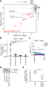
Transcription factor RUNX1 drives RASGRP1 expression, and is reduced in autoimmunity. (A) Affinity‐purification mass spectrometry analysis was performed to identify proteins from Jurkat lysates binding WT versus mutant (42 bp scramble) enhancer sequence. Indicated in red are factors preferentially bound to WT, in black are non‐significant changing binders. (B and C) RUNX1 mRNA levels, normalized to β2m, in CD4+ T cells isolated from JIA patients’ blood and during remission (JIA‐R (emission), N = 8, in three experiments of N = 3 or N = 2) or active inflammatory disease (JIA‐Active, N = 8, in three experiments of N = 3 or N = 2), or synovial fluid during active disease (SF, N = 3, one experiment), compared to healthy donors (HC, N = 6, two experiments). Three separate experiments were performed. Shown are averages ± SD. 2–dCT values of RUNX1, corrected for B2M. (C) Plot comparing RASGRP1 and RUNX1 mRNA 2–ddCT expression in each sample, connected by a line. (D) Western blot of RUNX1 and α‐tubulin (loading control) showing a representative example of RUNX1 knockdown upon RUNX1 shRNA transduction in Jurkat cells (N = 3 separate transduction experiments). (E) RASGRP1 mRNA, normalized to B2M expression in Jurkat, comparing cells transduced with RUNX1 targeting shRNA or non‐targeting (control) shRNA, relative to untransduced Jurkat cells (2–ddCT). N = 3 separate transduction experiments. Shown are averages ± SD. One‐way ANOVA statistical test was performed in (B), Holm's‐Sidak post‐test, and Two‐tailed T‐test was performed in (E), significance is indicated, * p < 0.05, ** p < 0.01.
Figure 5
Figure 5
Tight regulation of RASGRP1 expression in T cells is vital to prevent disease. Top: RASGRP1 transcription is regulated by enhancer 1 binding transcription factors (RUNX1/CBFB). T cell receptor signaling upon antigen/MHC binding induces activation of the RasGRP1 dimer, allowing membrane recruitment and binding to Ras. Next, RasGRP1 releases GDP bound to Ras, allowing GTP binding and active Ras‐MAPK signaling. Bottom: The level of RASGRP1 expression and resulting Ras‐MAPK signaling affects thymocyte selection and clinical outcome: Reduced RASGRP1 expression results in lower Ras‐MAPK signaling, thus reduced positive thymocyte selection signals, and can cause autoimmunity. Absence of RASGRP1 results in a complete loss of positive thymocyte selection and immune deficiency, while increased levels of RASGRP1 expression and Ras‐MAPK signaling have been shown in T‐ALL. Together, tight regulation of RasGRP1 expression is essential to maintain healthy T cells and prevent disease.

References

    1. Ksionda, O. , Limnander, A. and Roose, J. P. , RasGRP Ras guanine nucleotide exchange factors in cancer. Front. Biol. 2013. 8: 508–532. - PMC - PubMed
    1. Vercoulen, Y. , Kondo, Y. , Iwig, J. S. , Janssen, A. B. , White, K. A. , Amini, M. , Barber, D. L. et al., A Histidine pH sensor regulates activation of the Ras‐specific guanine nucleotide exchange factor RasGRP1. Elife 2017. 6. - PMC - PubMed
    1. Iwig, J. S. , Vercoulen, Y. , Das, R. , Barros, T. , Limnander, A. , Che, Y. , Pelton, J. G. et al., Structural analysis of autoinhibition in the Ras‐specific exchange factor RasGRP1. Elife 2013. 2: e00813. - PMC - PubMed
    1. Dower, N. A. , Stang, S. L. , Bottorff, D. A. , Ebinu, J. O. , Dickie, P. , Ostergaard, H. L. , Stone, J. C. , RasGRP is essential for mouse thymocyte differentiation and TCR signaling. Nat. Immunol. 2000. 1: 317–321. - PubMed
    1. Priatel, J. J. , Teh, S. J. , Dower, N. A. , Stone, J. C. and Teh, H. S. , RasGRP1 transduces low‐grade TCR signals which are critical for T cell development, homeostasis, and differentiation. Immunity 2002. 17: 617–627. - PubMed

Publication types

MeSH terms