. 2020 Oct 13;10(10):2014.
doi: 10.3390/nano10102014.

Magnetite-Arginine Nanoparticles as a Multifunctional Biomedical Tool

Affiliations

Magnetite-Arginine Nanoparticles as a Multifunctional Biomedical Tool

Victoria E Reichel et al. Nanomaterials (Basel). .

Abstract

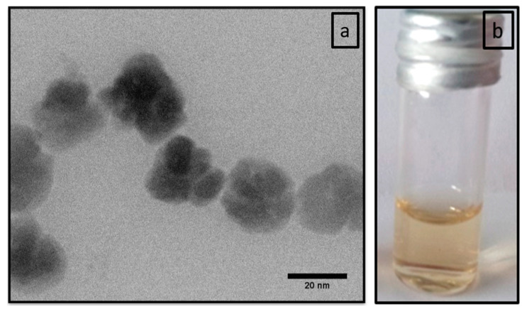
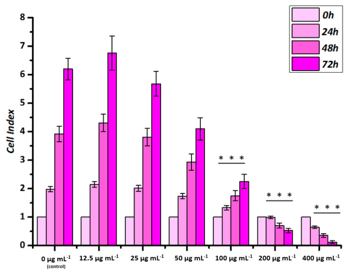
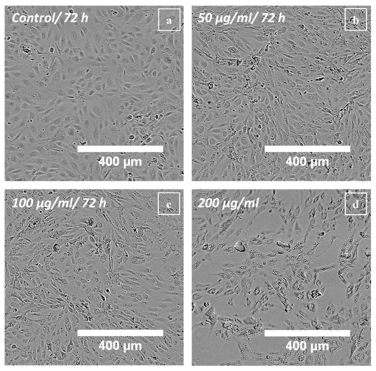
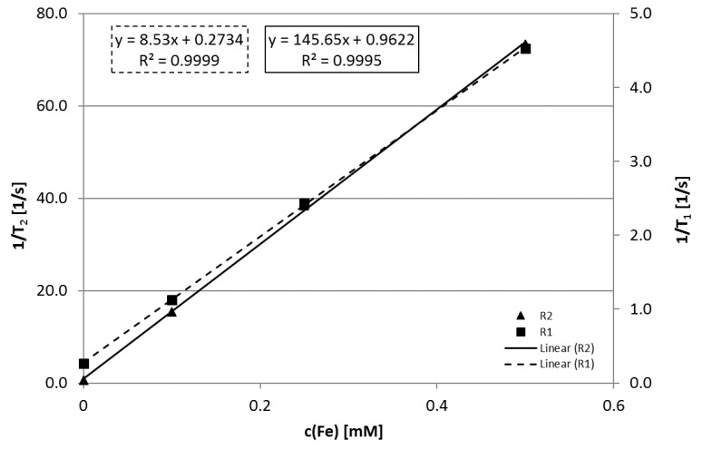
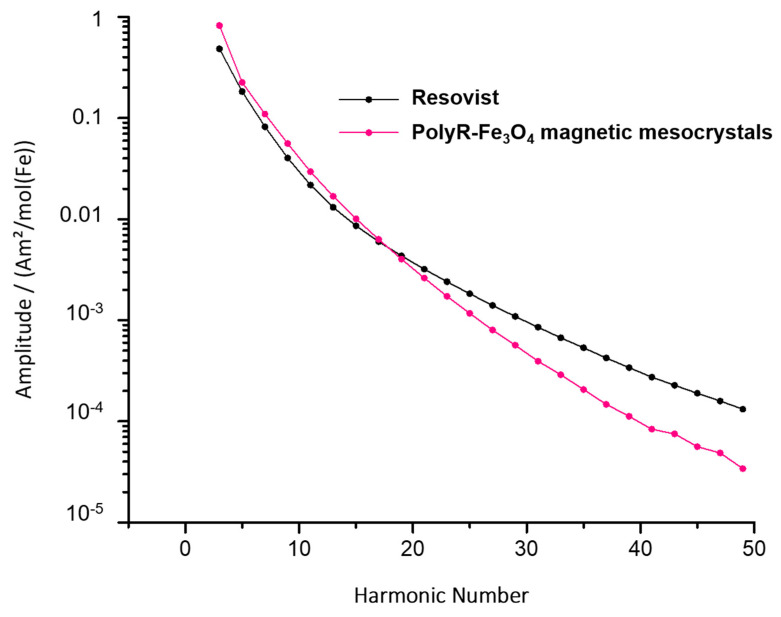
Iron oxide nanoparticles are a promising platform for biomedical applications, both in terms of diagnostics and therapeutics. In addition, arginine-rich polypeptides are known to penetrate across cell membranes. Here, we thus introduce a system based on magnetite nanoparticles and the polypeptide poly-l-arginine (polyR-Fe3O4). We show that the hybrid nanoparticles exhibit a low cytotoxicity that is comparable to Resovist®, a commercially available drug. PolyR-Fe3O4 particles perform very well in diagnostic applications, such as magnetic particle imaging (1.7 and 1.35 higher signal respectively for the 3rd and 11th harmonic when compared to Resovist®), or as contrast agents for magnetic resonance imaging (R2/R1 ratio of 17 as compared to 11 at 0.94 T for Resovist®). Moreover, these novel particles can also be used for therapeutic purposes such as hyperthermia, achieving a specific heating power ratio of 208 W/g as compared to 83 W/g for Feridex®, another commercially available product. Therefore, we envision such materials to play a role in the future theranostic applications, where the arginine ability to deliver cargo into the cell can be coupled to the magnetite imaging properties and cancer fighting activity.

Keywords: MRI; hyperthermia; iron oxide; nanoparticle; theranostics.


Conflict of interest statement

The authors declare no conflict of interest.

Figures

Figure 1
Figure 1
(a) STEM image of polyR-Fe3O4 nanoparticles. (b) Image of vial with colloidal stable polyR-Fe3O4 nanoparticles solution one year after synthesis.
Figure 2
Figure 2
Intensity-weighted log-normal distribution of the hydrodynamic diameter of polyR-Fe3O4 at different time points after synthesis. The mean hydrodynamic diameter d¯ is displayed for comparison.
Figure 3
Figure 3
Biological effects of polyR-Fe3O4 particles on endothelial cells grown in static conditions analyzed by real-time cell analysis. HUVECs were seeded 24 h before nanoparticle application. After the initial 24 h, particles at different concentrations were added and cell index was monitored for up to 72 h post application. Cell Index is displayed as x-fold of untreated controls. Data are expressed as mean ± SEM of n = 3 experiments performed in hexaplicate, *** p < 0.001 vs. corresponding control (One-way Anova).
Figure 4
Figure 4
Live-cell microscopy of HUVECs treated with polyR-Fe3O4: Phase contrast images of HUVECs were taken after 72 h post particle addition. (a) Untreated control HUVECs, (b) HUVECs treated with 50 µg mL−1, (c) HUVECs treated with 100 µg mL−1 and (d) HUVECs treated with 200 µg mL−1 polyR-Fe3O4. Representative images of n = 3 experiments and hexaplicate samples are shown.
Figure 5
Figure 5
Effects of circulating polyR-Fe3O4 on enthodelial cell grown under flow conditions. HUVECs were grown in bifurcating slides until confluence and perfused with polyR-Fe3O4-containing medium for 18 h. The representative laminar and non-uniform regions are shown after fluorescent staining. Nuclei are visualized using a Hoechst 33342 (blue) staining, whereas F-actin is visualized with Alexa 488-conjugated phalloidin (green). Representative images of n = 3 experiments are shown. (Scale bars: 50 μm).
Figure 6
Figure 6
Relationship of the iron concentration c(Fe) in [mM] and the reciprocal of the relaxation time 1/T1,2 in [1/s]. The relaxivities R1 and R2 were calculated using the slope of the obtained linear equation.
Figure 7
Figure 7
MPS measurement of polyR-Fe3O4 particles (pink) and Resovist® (black) as comparison.

References

    1. Silva A.K., Espinosa A., Wilhelm C., Gazeau F., Kolosnjaj-Tabi J. Medical Applications of Iron Oxide Nanoparticles. Iron Oxides. 2016:425–472. doi: 10.1002/9783527691395.ch18. - DOI
    1. Hergt R., Dutz S. Magnetic particle hyperthermia—Biophysical limitations of a visionary tumour therapy. J. Magn. Magn. Mater. 2007;311:187–192. doi: 10.1016/j.jmmm.2006.10.1156. - DOI
    1. Reichel V., Faivre D. New Perspectives on Mineral Nucleation and Growth. Springer International Publishing; Cham, Switzerland: 2017. Magnetite Nucleation and Growth; pp. 275–291. - DOI
    1. Yu E.Y., Bishop M., Zheng B., Ferguson R.M., Khandhar A.P., Kemp S.J., Krishnan K.M., Goodwill P.W., Conolly S.M. Magnetic Particle Imaging: A Novel in Vivo Imaging Platform for Cancer Detection. Nano Lett. 2017;17:1648–1654. doi: 10.1021/acs.nanolett.6b04865. - DOI - PMC - PubMed
    1. Shakeri S., Ashrafizadeh M., Zarrabi A., Roghanian R., Afshar E.G., Pardakhty A., Mohammadinejad R., Kumar A., Thakur V.K. Multifunctional Polymeric Nanoplatforms for Brain Diseases Diagnosis, Therapy and Theranostics. Biomedicines. 2020;8:13. doi: 10.3390/biomedicines8010013. - DOI - PMC - PubMed

LinkOut - more resources