Review
. 2020 Oct 13;10(10):1437.
doi: 10.3390/biom10101437.

The Dichotomous Role of Inflammation in the CNS: A Mitochondrial Point of View

Affiliations
Review

The Dichotomous Role of Inflammation in the CNS: A Mitochondrial Point of View

Bianca Vezzani et al. Biomolecules. .

Abstract

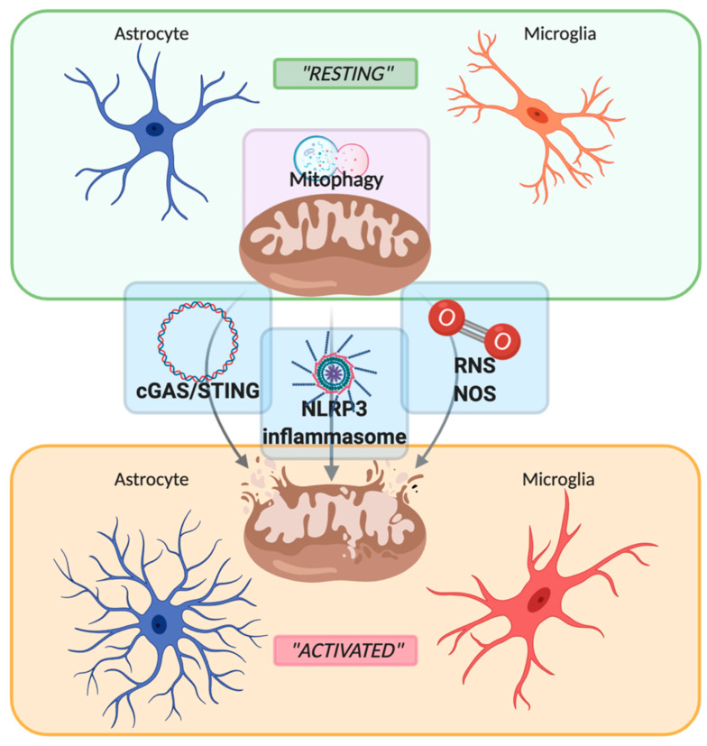
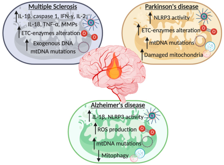
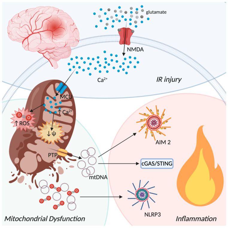
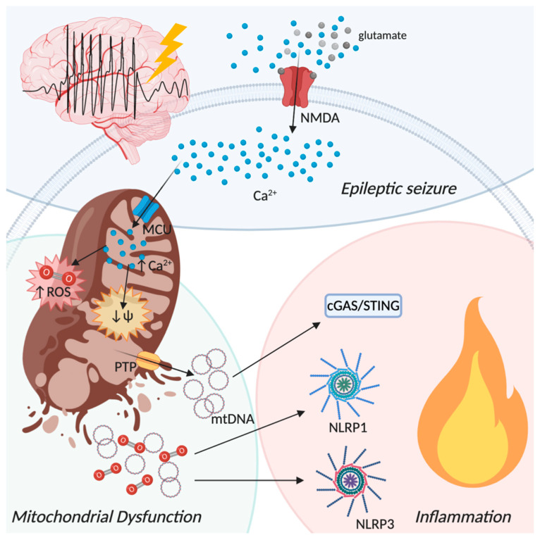
Innate immune response is one of our primary defenses against pathogens infection, although, if dysregulated, it represents the leading cause of chronic tissue inflammation. This dualism is even more present in the central nervous system, where neuroinflammation is both important for the activation of reparatory mechanisms and, at the same time, leads to the release of detrimental factors that induce neurons loss. Key players in modulating the neuroinflammatory response are mitochondria. Indeed, they are responsible for a variety of cell mechanisms that control tissue homeostasis, such as autophagy, apoptosis, energy production, and also inflammation. Accordingly, it is widely recognized that mitochondria exert a pivotal role in the development of neurodegenerative diseases, such as multiple sclerosis, Parkinson's and Alzheimer's diseases, as well as in acute brain damage, such in ischemic stroke and epileptic seizures. In this review, we will describe the role of mitochondria molecular signaling in regulating neuroinflammation in central nervous system (CNS) diseases, by focusing on pattern recognition receptors (PRRs) signaling, reactive oxygen species (ROS) production, and mitophagy, giving a hint on the possible therapeutic approaches targeting mitochondrial pathways involved in inflammation.

Keywords: Alzheimer’s disease; Parkinson’s disease; epilepsy; ischemic stroke; mitochondria; multiple sclerosis; neurodegeneration; neuroinflammation.


Conflict of interest statement

The authors declare no conflict of interest.

Figures

Figure 1
Figure 1
Schematic representation of the switch between “resting” and “activated” state of astrocyte and microglia mediated by mitochondria. Mitochondria maintain their healthy and physiological state by mitophagy. Upon stressful condition, such as inflammatory stimuli, mitochondria are disrupted with the consequent release of mtDNA, damage-associated molecular patterns (DAMPs), reactive oxygen/nitrogen species (ROS and RNS), leading to the activation of the sentinel of the central nervous system such as astrocytes and microglia. Created with BioRender.com.
Figure 2
Figure 2
Schematic list of mitochondrial contribution to the recurrence of neuroinflammation during the development of neurodegenerative diseases (NDDs). In multiple sclerosis, Parkinson’s disease, and Alzheimer’s disease mitochondria have a fundamental role in the induction of a neuroinflammatory state by the activation of inflammasomes, variation of the electron transport chain (ETC) enzymes, modulation of reactive oxygen species (ROS) production, alteration of the mtDNA and of the mitophagic process. Created with BioRender.com.
Figure 3
Figure 3
Ischemic-reperfusion injury from a mitochondrial perspective. Oxygen depletion occurring during an ischemic event lead to neuronal membrane depolarization with glutamate release in the extracellular space. Glutamate binds to N-methyl-D-aspartate (NMDA) receptors promoting a strong influx of Ca2+. The increase of cytosolic Ca2+ activates the mitochondria calcium uniporter (MCU) leading to mitochondrial depolarization and increased production of reactive oxygen species (ROS) with the consequent opening of the permeability transition pore (PTP) and the final disruption of the mitochondrial membranes. mtDNA and ROS release in the cytoplasm drives the inflammatory process by the activation different pathways, such as AIM2 and the NLRP3 inflammasomes and the cGAS-STING dsDNA sensing machinery. Created with BioRender.com.
Figure 4
Figure 4
Epileptic seizure from a mitochondrial perspective. Repeated epileptic seizures activate NMDA receptors with the consequent influx of Ca2+ into the cytoplasm. Additionally, in this scenario, increased cytoplasmic Ca2+ concentration activates MCU. Augmented mitochondrial Ca2+ level triggers the activation of different enzymes responsible for reactive oxygen species production (ROS), such as nitric-oxide synthase and NADPH oxidase, and leads to membrane depolarization with the opening of the permeability transition pore (PTP) and mitochondrial membrane disruption. Release of mitochondrial components in the cytoplasm activates proinflammatory pathways such as NLRP1 and NLRP3 inflammasome assembly and the cGAS-STING dsDNA sensing machinery, leading to progressive inflammation. Created with BioRender.com.

References

    1. Barker C.F., Billingham R.E. Immunologically privileged sites. Adv. Immunol. 1977;25:1–54. - PubMed
    1. Medawar P.B. Immunity to homologous grafted skin; the fate of skin homografts transplanted to the brain, to subcutaneous tissue, and to the anterior chamber of the eye. Br. J. Exp. Pathol. 1948;29:58–69. - PMC - PubMed
    1. Louveau A., Harris T.H., Kipnis J. Revisiting the Mechanisms of CNS Immune Privilege. Trends Immunol. 2015;36:569–577. doi: 10.1016/j.it.2015.08.006. - DOI - PMC - PubMed
    1. Louveau A., Smirnov I., Keyes T.J., Eccles J.D., Rouhani S.J., Peske J.D., Derecki N.C., Castle D., Mandell J.W., Lee K.S., et al. Structural and functional features of central nervous system lymphatic vessels. Nature. 2015;523:337–341. doi: 10.1038/nature14432. - DOI - PMC - PubMed
    1. Aspelund A., Antila S., Proulx S.T., Karlsen T.V., Karaman S., Detmar M., Wiig H., Alitalo K. A dural lymphatic vascular system that drains brain interstitial fluid and macromolecules. J. Exp. Med. 2015;212:991–999. doi: 10.1084/jem.20142290. - DOI - PMC - PubMed

Publication types

MeSH terms