. 2020 Oct 13;10(10):726.
doi: 10.3390/brainsci10100726.

Prediction of Human Inhibition Brain Function with Inter-Subject and Intra-Subject Variability

Affiliations

Prediction of Human Inhibition Brain Function with Inter-Subject and Intra-Subject Variability

Rupesh Kumar Chikara et al. Brain Sci. .

Abstract

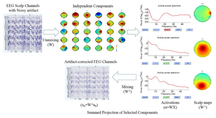
The stop signal task has been used to quantify the human inhibitory control. The inter-subject and intra-subject variability was investigated under the inhibition of human response with a realistic environmental scenario. In present study, we used a battleground scenario where a sniper-scope picture was the background, a target picture was a go signal, and a nontarget picture was a stop signal. The task instructions were to respond on the target image and inhibit the response if a nontarget image appeared. This scenario produced a threatening situation and endorsed the evaluation of how subject's response inhibition manifests in a real situation. In this study, 32 channels of electroencephalography (EEG) signals were collected from 20 participants during successful stop (response inhibition) and failed stop (response) trials. These EEG signals were used to predict two possible outcomes: successful stop or failed stop. The inter-subject variability (between-subjects) and intra-subject variability (within-subjects) affect the performance of participants in the classification system. The EEG signals of successful stop versus failed stop trials were classified using quadratic discriminant analysis (QDA) and linear discriminant analysis (LDA) (i.e., parametric) and K-nearest neighbor classifier (KNNC) and Parzen density-based (PARZEN) (i.e., nonparametric) under inter- and intra-subject variability. The EEG activities were found to increase during response inhibition in the frontal cortex (F3 and F4), presupplementary motor area (C3 and C4), parietal lobe (P3 and P4), and occipital (O1 and O2) lobe. Therefore, power spectral density (PSD) of EEG signals (1-50Hz) in F3, F4, C3, C4, P3, P4, O1, and O2 electrodes were measured in successful stop and failed stop trials. The PSD of the EEG signals was used as the feature input for the classifiers. Our proposed method shows an intra-subject classification accuracy of 97.61% for subject 15 with QDA classifier in C3 (left motor cortex) and an overall inter-subject classification accuracy of 71.66% ± 9.81% with the KNNC classifier in F3 (left frontal lobe). These results display how inter-subject and intra-subject variability affects the performance of the classification system. These findings can be used effectively to improve the psychopathology of attention deficit hyperactivity disorder (ADHD), obsessive-compulsive disorder (OCD), schizophrenia, and suicidality.

Keywords: classification; electroencephalography; frontal cortex; inter-subject variability; intra-subject variability; machine learning; prediction; response inhibition.


Conflict of interest statement

The authors declare no conflict of interest.

Figures

Figure 1
Figure 1
Design of stop signal task: (A) go trial and (B) stop trial under battleground scenario. Each subject performed go trials (75%) and stop trials (25%) according to the stimuli presented in the battleground scenario. (C) The theoretical model of the stop-signal task and where P is the probability of responding to the stop-signal.
Figure 2
Figure 2
The electroencephalography (EEG) devices used in battleground scenario. (A) Neuro Scan NuAmps signal amplifier. (B) EEG cap with 32-channels. (C) Experimental screen in battleground scenario.
Figure 3
Figure 3
Flowchart of the independent component analysis and back projecting the retained independent components to artifact free EEG channels (i.e., clean EEG signals).
Figure 4
Figure 4
Flowchart of the classification system architecture. (I). Preprocessing steps of acquired EEG signals. (II). Human inhibition-related brain regions. (III). Analysis of EEG signals power spectral density (PSD) under inter-subject and intra-subject at preparation state before stimulus. (IV). The QDA and LDA (parametric) and KNNC and PARZEN (nonparametric) classifiers performance outcomes comparison during inter-subject and intra-subject variability.
Figure 5
Figure 5
The average event-related potential (ERP) during go, successful stop, and failed stop trials at F3, F4, and C3 channels. Yellow asterisks indicate pairwise significance (p < 0.01) in Wilcoxon signed-rank test between the go and successful stop conditions. Green asterisks show pairwise significance (p < 0.01) in Wilcoxon signed-rank test between the go and failed stop. Violet asterisks show pairwise significance (p < 0.01) in Wilcoxon signed-rank test between the successful and failed stop.
Figure 6
Figure 6
The average event-related spectral perturbation (ERSP) of the frontal cortex and the supplementary motor area of the brain during go, successful stop, and failed stop trials at F3, F4, and C3 channels. The first magenta line shows go-stimulus onset. The second black dashed line reveals stop signal onset. The third black dashed line presents response onset. Statistically significant at p < 0.01. Color bars show the scale of ERSP.
Figure 7
Figure 7
The average ERSP of the EEG signals under “Successful stop—Go” and “Failed stop—Go” conditions at F3, F4, and C3 channels. The first magenta line shows go-stimulus onset. The second black dashed line reveals stop signal onset. The third black dashed line presents response onset. Statistically significant at p < 0.01. Color bars show the scale of ERSP.
Figure 8
Figure 8
The average power spectral density (PSD) of all subjects (inter-subject variability) at F3, F4, C3, C4, P3, P4, O1, and O2 under successful stop and failed stop trials. Asterisk show significant difference between successful stop trials and failed stop trials by Wilcoxon signed-rank test (at *p < 0.05).
Figure 9
Figure 9
The QDA and LDA (parametric) and KNNC and PARZEN (nonparametric) classifiers performance outcomes comparison in accuracy and standard error bars during inter-subject and intra-subject variability at F3, F4, C3, C4, P3, P4, O1, and O2. Asterisks show pairwise significance difference (* p < 0.05) in t-test between the QDA, LDA, KNNC, and PARZEN.

Similar articles

Cited by

References

    1. Aron A.R. The neural basis of inhibition in cognitive control. Neuroscientist. 2007;13:214–228. doi: 10.1177/1073858407299288. - DOI - PubMed
    1. Chambers C.D., Garavan H., Bellgrove M.A. Insights into the neural basis of response inhibition from cognitive and clinical neuroscience. Neurosci. Biobehav. Rev. 2009;33:631–646. doi: 10.1016/j.neubiorev.2008.08.016. - DOI - PubMed
    1. Verbruggen F., Logan G.D. Response inhibition in the stop-signal paradigm. Trends Cogn. Sci. 2008;12:11418–11424. - PMC - PubMed
    1. Verbruggen F., Logan G.D. Models of response inhibition in the stop-signal and stop-change paradigms. Neurosci. Biobehav. Rev. 2009;33:647–661. doi: 10.1016/j.neubiorev.2008.08.014. - DOI - PMC - PubMed
    1. Lappin J., Eriksen C. Use of delayed signal to stop a visual reaction-time response. J. Exp. Psychol. 1966;72:805–811. doi: 10.1037/h0021266. - DOI

LinkOut - more resources