. 2020 Oct 13;8(10):413.
doi: 10.3390/biomedicines8100413.

Preliminary Study on β3-Adrenoreceptor as Predictor Marker of Relapse in Ewing Sarcoma Patients

Affiliations

Preliminary Study on β3-Adrenoreceptor as Predictor Marker of Relapse in Ewing Sarcoma Patients

Maura Calvani et al. Biomedicines. .

Abstract

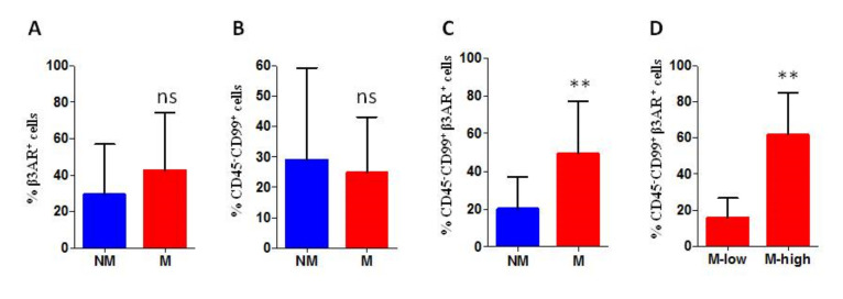
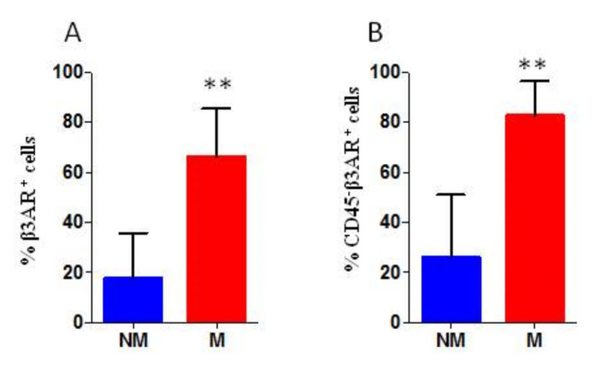
Ewing sarcoma (EWS) is a paediatric aggressive malignant tumour of bones and soft tissues. Multidisciplinary chemotherapies, surgical resection, and radiation represent the only strategies counteracting the disease, however spreading and relapse of disease still remain a clinical issue. Circulating tumour cells (CTCs) are an important feature of EWS but the prognostic significance has not been, yet, clarified. CTCs have been found both in patients with localized disease and in those who recur or metastasize. The identification of markers that can detect recurrences and metastasis remains an important challenge for research. Unfortunately, even most of patients with localized cancer relapsed and the reason has not yet been fully understood. In this clinical study on EWS patients, we evaluated the expression of CD99 antigen and beta-3 adrenergic receptor (β3-AR) on CTCs and bioptic derived cells by flow cytometry. The preliminary data revealed a higher β3-AR expression on cells derived from metastatic or relapsed patients, suggesting a role for the β3-AR as a possible predictive maker of disease recurrence in both patients with metastatic and localized disease.

Keywords: circulating tumour cells; ewing sarcoma; β3-adrenergic receptors.


Conflict of interest statement

The authors declare no conflict of interest.

Figures

Figure 1
Figure 1
The figure represents the gating strategy used for the cytofluorimetric analysis of all samples.
Figure 2
Figure 2
Cytofluorimetric (FACS) analysis of peripheral blood derived cells, the percentage of positive cells is reported. (A) percentage of beta-3-adrenergic receptor (β3-AR) positive cells within of all peripheral blood (PB) cells (% β3-AR+ cells range: Non-metastatic (NM) 11–58%; metastatic (M) 25–75%); (B) percentage of CD99 positive cells on the surface of CD45 PB cells (% CD99+ cells range: NM 0–59%; M 8–43%); (C) percentage of β3-AR positive cells on CD45 CD99+ gated cells (% β3-AR+ cells range: NM 0–39%; M 5–80%); and (D) percentage of β3-AR positive cells on CD45 CD99+ gated cells analysed within the group of metastatic patients (% β3-AR+ cells range: Mhigh 37–80%; Mlow 5–23%). M low: M group with low β3-AR expression level, and M high: M group with high β3-AR expression level. PB from 20 patients: 10 M and 10 NM. ns: Not significant. p values for analysis ** p < 0.01.
Figure 3
Figure 3
Kaplan Meier curve analysis showing the progression free survival (PFS) in metastatic patients divided in the two subgroups of high and low β3-AR expression. The curve shows the positive association between high β3-AR levels and progression of the disease. High: Metastatic patients with percentage of β3-AR positive cells higher than 30%; Low: Metastatic patients with percentage of β3-AR positive cells lower than 30%.
Figure 4
Figure 4
Cytofluorimetric analysis of bone marrow derived cells, the percentage of positive cells is reported. (A) percentage of β3-AR positive cells within all BM cells (% β3-AR+ cells range: NM 2.5–48%; M 46–85%) and (B) percentage of β3-AR positive cells within CD45 gated BM cells (% β3-AR+ cells range: NM 0.5–56%; M 65–91%). BM from 14 patients: 5 M and 9 NM. P values for analysis: ** p < 0.01.
Figure 5
Figure 5
Cytofluorimetric analysis of biopsies derived cells, the percentage of positive cells is reported. (A) percentage of β3-AR positive cells within all cells derived from the biopsy (% β3-AR+ cells range: NM 6–44%; M 43–87%); (B) percentage of β3-AR positive cells within CD99+ gated cancer cells (% β3-AR+ cells range: NM 18–53%; M 63–86%); and (C) percentage of β3-AR positive cells within CD45 gated cells (% β3-AR+ cells range: NM 4–39%; M 43–84%). Biopsy samples from 7 patients: 4 M and 3 NM. p values for analysis: * p < 0.05 and ** p < 0.01.
Figure 6
Figure 6
In vitro analysis of CD99 expression after β3-ARs blockade. (A) analysis of cellular viability with MTT survival experiment in A673 and SK-ES-1 cell lines and (B) CD99 mean fluorescence intensity (MFI) in A673 and SK-ES-1 cells investigated by cytofluorimetric analysis, after 24 h-treatment with SR59230A 5 M (SR5uM) and 10 M (SR10uM); ctrl: Untreated.
Figure 7
Figure 7
Schematic representation of the preliminary results on metastatic patients (left) and the suggested evaluation during the follow-up even for localized patients (right).
Figure 8
Figure 8
The figure represents in blue squares the routinely workflow for diagnosis and in the coloured squares the new prognostic workflow suggested according to our preliminary results.

Similar articles

Cited by

References

    1. Dasgupta A., Trucco M., Rainusso N., Bernardi R.J., Shuck R., Kurenbekova L., Loeb D.M., Yustein J.T. Metabolic modulation of Ewing sarcoma cells inhibits tumor growth and stem cell properties. Oncotarget. 2017;8:77292–77308. doi: 10.18632/oncotarget.20467. - DOI - PMC - PubMed
    1. Hoang N.T., Acevedo L.A., Mann M.J., Tolani B. A review of soft-tissue sarcomas: Translation of biological advances into treatment measures. Cancer Manag. Res. 2018;10:1089–1114. doi: 10.2147/CMAR.S159641. - DOI - PMC - PubMed
    1. Gaspar N., Hawkins D.S., Dirksen U., Lewis I.J., Ferrari S., Le Deley M.-C., Kovar H., Grimer R.J., Whelan J., Claude L., et al. Ewing Sarcoma: Current Management and Future Approaches Through Collaboration. J. Clin. Oncol. 2015;33:3036–3046. doi: 10.1200/JCO.2014.59.5256. - DOI - PubMed
    1. Riggi N., Stamenkovic I. The Biology of Ewing sarcoma. Cancer Lett. 2007;254:1–10. doi: 10.1016/j.canlet.2006.12.009. - DOI - PubMed
    1. Karski E.E., Matthay K.K., Neuhaus J.M., Goldsby R.E., Dubois S.G. Characteristics and outcomes of patients with Ewing sarcoma over 40 years of age at diagnosis. Cancer Epidemiol. 2013;37:29–33. doi: 10.1016/j.canep.2012.08.006. - DOI - PMC - PubMed