Review
. 2020 Oct 13;9(4):53.
doi: 10.3390/antib9040053.

Structure, Function, and Therapeutic Use of IgM Antibodies

Affiliations
Review

Structure, Function, and Therapeutic Use of IgM Antibodies

Bruce A Keyt et al. Antibodies (Basel). .

Abstract

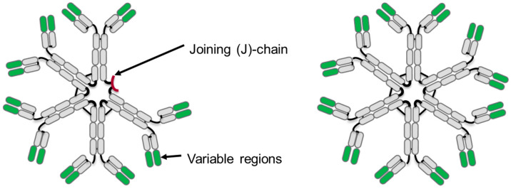
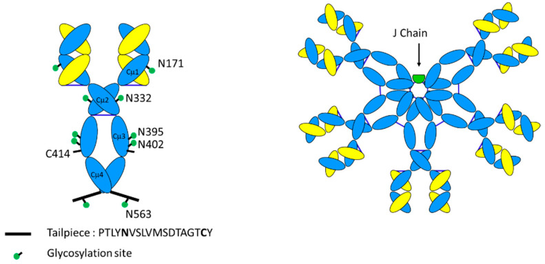
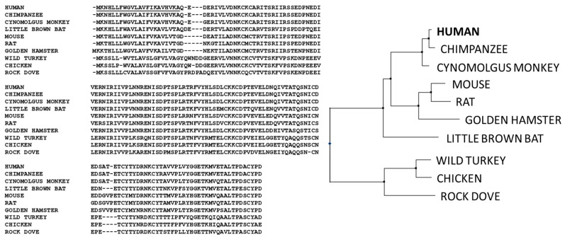
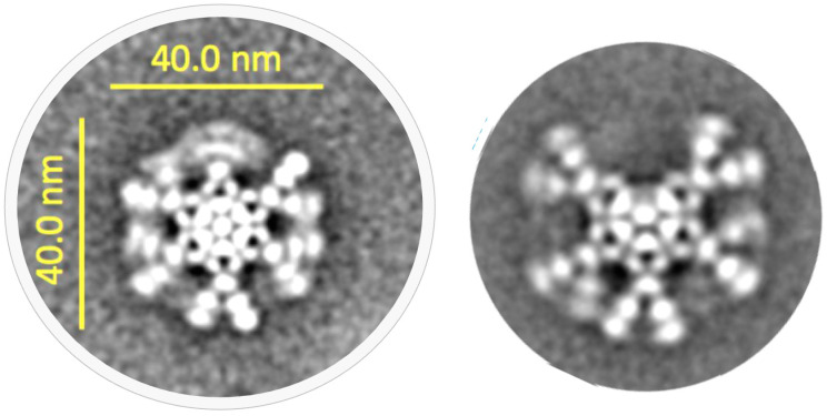
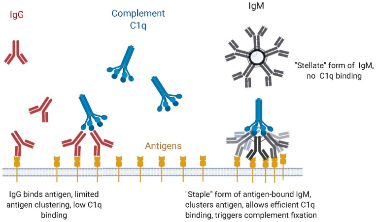
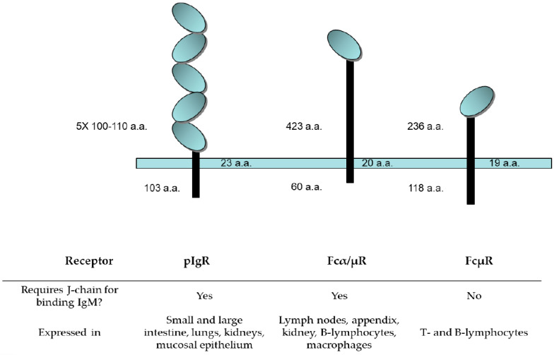
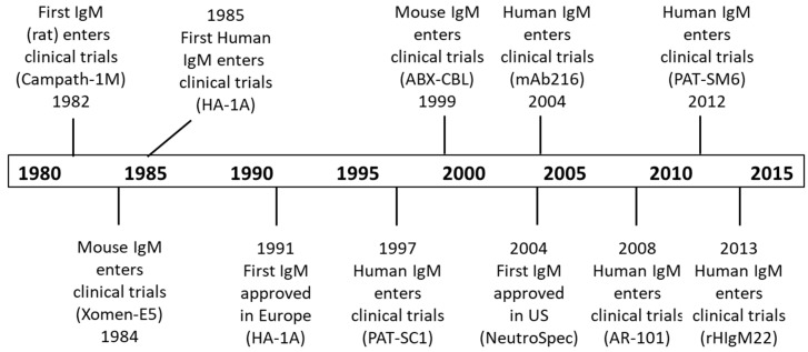
Natural immunoglobulin M (IgM) antibodies are pentameric or hexameric macro-immunoglobulins and have been highly conserved during evolution. IgMs are initially expressed during B cell ontogeny and are the first antibodies secreted following exposure to foreign antigens. The IgM multimer has either 10 (pentamer) or 12 (hexamer) antigen binding domains consisting of paired µ heavy chains with four constant domains, each with a single variable domain, paired with a corresponding light chain. Although the antigen binding affinities of natural IgM antibodies are typically lower than IgG, their polyvalency allows for high avidity binding and efficient engagement of complement to induce complement-dependent cell lysis. The high avidity of IgM antibodies renders them particularly efficient at binding antigens present at low levels, and non-protein antigens, for example, carbohydrates or lipids present on microbial surfaces. Pentameric IgM antibodies also contain a joining (J) chain that stabilizes the pentameric structure and enables binding to several receptors. One such receptor, the polymeric immunoglobulin receptor (pIgR), is responsible for transcytosis from the vasculature to the mucosal surfaces of the lung and gastrointestinal tract. Several naturally occurring IgM antibodies have been explored as therapeutics in clinical trials, and a new class of molecules, engineered IgM antibodies with enhanced binding and/or additional functional properties are being evaluated in humans. Here, we review the considerable progress that has been made regarding the understanding of biology, structure, function, manufacturing, and therapeutic potential of IgM antibodies since their discovery more than 80 years ago.

Keywords: IgM (immunoglobulin M); avidity; complement dependent cytotoxicity (CDC); hexameric; joining chain (J-chain); pentameric; poly Ig receptor (pIgR); polymeric; polyvalency.


Conflict of interest statement

The authors declare the following potential conflict of interest: B.A.K., A.M.S., S.F.C., and M.S.P. are currently employees of IGM Biosciences and hold stock in that company. R.B. was an employee of IGM Biosciences and holds stock in that company. The funders had no role in the design of the study; in the collection, analyses, or interpretation of data; in the writing of the manuscript, or in the decision to publish the results.

Figures

Figure 1
Figure 1
Schematic diagram of an immunoglobulin M (IgM) antibody pentamer (left) and hexamer (right). Constant regions are shown in gray and variable regions in green, and also shown on the IgM pentamer is the small joining chain (J-chain) in red.
Figure 2
Figure 2
Schematic diagram illustrating the evolution of immunoglobin (Ig) heavy and light chain isotypes in vertebrates, with the IgM isotype broadly represented across phyla. Antigen-binding variable lymphocyte receptors (VLRs) in jawless fishes (agnathans) are thought to be precursors of immunoglobulins. The IgW isotype in cartilaginous fishes is orthologous to IgD in other groups; IgNAR is a “new antigen receptor” isotype, identified in nurse shark, that does not associate with light chains and does not have an ortholog in higher species. IgT appears to be the most ancient Ig specialized for mucosal protection. IgX, originally identified in Xenopus is orthologous and functionally analogous to IgA. IgY is the amphibian, reptilian, and avian equivalent of IgG and IgE. IgF only has two constant domains but has homology to IgY. Open boxes represent the lack of certain heavy or light chains in the certain vertebrate lineages; dashed boxes represent a common ancestry; and * represents the lack of κ light chain in snakes. Figure adapted from Pettinello and Dooley 2014 [22] and Kaetzel 2014 [23].
Figure 3
Figure 3
Sequence alignment of the heavy chains of different isotypes of immunoglobulin showing the glycosylation sites and location of inter- and intra-disulfide linkages. The Cμ2 region of IgMs and IgEs is analogous to the hinge regions of the other isotypes. The alignments also highlight, in bold font, the locations of glycosylation sites on each heavy chain. Sequences are numbered according the convention established by Kabat [74].
Figure 3
Figure 3
Sequence alignment of the heavy chains of different isotypes of immunoglobulin showing the glycosylation sites and location of inter- and intra-disulfide linkages. The Cμ2 region of IgMs and IgEs is analogous to the hinge regions of the other isotypes. The alignments also highlight, in bold font, the locations of glycosylation sites on each heavy chain. Sequences are numbered according the convention established by Kabat [74].
Figure 4
Figure 4
Schematic diagram of IgM monomer vs. IgM pentamer. IgM monomers are distinguished from IgG counterparts by their extensive glycosylation at the asparagine residues indicated, the presence of an additional domain Cµ2 in place of a hinge, and the presence of a short tailpiece peptide sequence that is critical for multimerization. Pentameric IgM has an additional 137 amino acid joining (J)-chain.
Figure 5
Figure 5
Sequence alignment (left) and hierarchical clustering (CLUSTALW, right) of IgM joining chains (J-chains) showing the high degree of conservation across species from human, primates, rodents, and birds.
Figure 6
Figure 6
Example cryo-electron microscopy (cryo-EM) results for hexamer (left) and pentamer (right) forms of an anti-CD20 IgM. Consistent with published observations [91], the pentamer formed in the absence of J-chain retains the positions of monomers around the central ring and only a single monomer appears substituted by the J-chain. These images are a montage of a large series of negatively stained, transmission electron micrographs obtained via collaboration of IGM Biosciences and NanoImaging, Inc., San Diego, CA, USA (unpublished results).
Figure 7
Figure 7
Complement binding and activation with IgM as compared with IgG.
Figure 8
Figure 8
Schematic diagram of receptors known to bind IgM. IgMs bind at least three different receptors from those that bind IgG. The oval domains of each receptor indicate immunoglobulin fold-like regions. Their sizes and tissue distribution are depicted above.
Figure 9
Figure 9
History of selected IgM human clinical trials.

References

    1. Nuttall G. Experimente uber die bacterienfeindlichen Einflusse des theirischen Korpers. Z. Hyg. Infektionskr. 1888;4:353–395.
    1. Black C.A. A brief history of the discovery of the immunoglobulins and the origin of the modern immunoglobulin nomenclature. Immunol. Cell Biol. 1997;75:65–68. doi: 10.1038/icb.1997.10. - DOI - PubMed
    1. Kabat E.A. The Molecular Weight of Antibodies. J. Exp. Med. 1939;69:103–118. doi: 10.1084/jem.69.1.103. - DOI - PMC - PubMed
    1. Waldenstrom J. Incipient myelomatisis or “essential” hyperglobulinemia with fibrinogenopenia—A new syndrome? Acta Med. Scand. 1944;67:216–247.
    1. Wallenius G., Trautman R., Kunkel H.G., Franklin E.C. Ultracentrifugal studies of major non-lipide electrophoretic components of normal human serum. J. Biol. Chem. 1957;225:253–267. - PubMed

LinkOut - more resources