. 2020 Oct 14;9(10):2289.
doi: 10.3390/cells9102289.

Cytotoxicity and Mitochondrial Dysregulation Caused by α-Synuclein in Dictyostelium discoideum

Affiliations

Cytotoxicity and Mitochondrial Dysregulation Caused by α-Synuclein in Dictyostelium discoideum

Sanjanie Fernando et al. Cells. .

Abstract

Alpha synuclein has been linked to both sporadic and familial forms of Parkinson's disease (PD) and is the most abundant protein in Lewy bodies a hallmark of Parkinson's disease. The function of this protein and the molecular mechanisms underlying its toxicity are still unclear, but many studies have suggested that the mechanism of α-synuclein toxicity involves alterations to mitochondrial function. Here we expressed human α-synuclein and two PD-causing α-synuclein mutant proteins (with a point mutation, A53T, and a C-terminal 20 amino acid truncation) in the eukaryotic model Dictyostelium discoideum. Mitochondrial disease has been well studied in D. discoideum and, unlike in mammals, mitochondrial dysfunction results in a clear set of defective phenotypes. These defective phenotypes are caused by the chronic hyperactivation of the cellular energy sensor, AMP-activated protein kinase (AMPK). Expression of α-synuclein wild type and mutant forms was toxic to the cells and mitochondrial function was dysregulated. Some but not all of the defective phenotypes could be rescued by down regulation of AMPK revealing both AMPK-dependent and -independent mechanisms. Importantly, we also show that the C-terminus of α-synuclein is required and sufficient for the localisation of the protein to the cell cortex in D. discoideum.

Keywords: AMPK; Dictyostelium; mitochondria; α-synuclein.


Conflict of interest statement

The authors declare no conflict of interest.

Figures
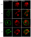
Figure A1
Figure A1
α-synuclein does not colocalise with mitochondria.
Figure 1
Figure 1
Human α-synuclein can be expressed in D. discoideum. Western blot showing the expression of α-synuclein WT HPF885 (38) and HPF891(96), α-synuclein A53T HPF899 (88) and HPF902(26) which runs at 18 kDa and α-synucleinΔC HPF910 (57) and HPF911(53) which runs at 14.5 kDa. The numbers in brackets represent construct copy numbers. Β-actin was detected as an indicator of sample load. The parental strain AX2 was run as a negative control.
Figure 2
Figure 2
In Dictyostelium, α-synuclein concentrates in the cell cortex and the C-terminus is necessary and sufficient for this. Immunofluorescent images of strains expressing various forms of α-synuclein. The α-synuclein was detected using an anti-α-synuclein antibody and visualized with an Alexa-Fluor-488-conjugated secondary antibody. DAPI was used to detect the nucleus. (a) WT α-synuclein expressed in the parental strain AX2 (strain HPF891) is enriched in the cortex of the cell. (b) Strain HPF899 expressing α-synuclein with the point mutation A53T also exhibited enrichment at the cortex, indicating that the subcellular localization is not affected by this mutation. (c) Strain HPF912 expressing the C terminal truncated mutant α-synuclein. The mutated protein appears granulated throughout the cytoplasm with no cortical signal observed. This indicates that the 20 residues of the C-terminal are necessary for enrichment of the protein in the cell cortex. (d) Cells expressing the C-terminal 20 amino acids of α-synuclein fused to GFP and visualized using an anti-GFP antibody. A strong signal is obvious at the cell cortex which suggests that these amino acids are sufficient for localization of α-synuclein to the cell cortex in Dictyostelium.
Figure 3
Figure 3
Mutant α-synuclein negatively affects phototaxis and thermotaxis. (a) Phototaxis. The parental strain (AX2), α-synuclein WT strain (HPF885), α-synuclein A53T mutant (HPF902) and α-synucleinΔC mutant (HPF905) were statistically analysed and the accuracy of phototaxis (κ) was plotted against their cell densities. When compared to AX2, the two mutant forms showed significantly decreased accuracy of phototaxis with the α-synucleinΔC mutant showing the lowest accuracy of phototaxis among the two forms. The α-synuclein WT strains showed a similar accuracy of phototaxis to AX2 at all cell densities. Error bars represent 90% confidence intervals. Lines were fitted with the least squares method to a logarithmic equation. (b) The accuracy of thermotaxis (κ) by slugs of the parental strain (AX2), and strains expressing the α-synuclein WT (HPF885), A53T point mutant (HPF902) and α-synucleinΔC mutant (HPF905) proteins was plotted against the temperature. The temperatures are expressed in arbitrary units from 1 to 8, shown in separate calibration experiments to correspond to agar surface temperatures of 14 °C to 28 °C at the centre of the plate [44]. When compared to AX2, strains expressing the α-synucleinΔC mutants show significantly reduced accuracy of thermotaxis while those expressing the α-synuclein WT or the α-synuclein A53T mutant form show a similar thermotaxis phenotype to AX2. Error bars represent 90% confidence intervals.
Figure 4
Figure 4
Multicellular morphogenesis of α-synuclein-expressing strains. Fruiting body morphology by the parental strain (AX2), strains expressing α-synuclein wild type (α-syn WT), point mutant (α-syn A53T), truncated (α-synΔC) α-synuclein and mitochondrial disease strain (Cpn60 inhibition). The α-syn WT and α-syn A53T produce thinner, longer stalks while the α-synΔC display fewer fruiting bodies with shorter and thicker stalks. Insets show the side view of a single fruiting body. The scale bar shows 1 mm.
Figure 5
Figure 5
α-synuclein affects growth on bacterial lawns but not in liquid media. (a) The plaque expansion rates by the α-synuclein-expressing strains plotted against the α-synuclein construct copy number. The tested strains were grown on E. coli B2 lawns on SM agar and the plaque expansion was measured over one hundred hours. Experiments were performed in triplicate in three individual experiments. Alpha-synuclein WT and the point mutant strains showed slower growth compared to the parental strain but were not significantly different from each other, the α-synucleinΔC strains showed an even slower growth compared to the parental strain. Error bars represent standard errors of the mean. The regression was significant at indicated p values (t-test). The lines were fitted by the least squares method to a linear equation. (b) The growth rate in liquid media of strains expressing α-synuclein was plotted against the α-synuclein construct copy number. The strains were grown in HL-5 medium in shaken cultures at 21 °C and the generation times (doubling time during exponential growth) were measured. Experiments were performed in duplicate in each of two separate experiments. Neither the α-synuclein WT nor the mutant forms caused adverse effects on growth in liquid. Error bars represent standard errors of the mean. The regression was not significant as indicated by the p value. The line was fitted by the least squares method to a linear equation.
Figure 6
Figure 6
α-Synuclein affects phagocytosis but not macropinocytosis. (a) Dictyostelium amoebae were fed E. coli expressing Ds-Red fluorescent protein and duplicate fluorescent measurements were taken immediately after the addition of bacteria and after 30 min of incubation at 21 °C on a shaker. The uptake rates were normalized to the uptake rate for AX2. The indicated number of strains (n) expressing each mutant α-synuclein form all showed a similar, significantly reduced rate of phagocytosis compared to the parental strain AX2, but there were no statistical differences among them (ANOVA with pairwise comparisons using the Least Squares Difference test). In a single sample t-test, the normalized phagocytosis rates for strains expressing α-synuclein forms were significantly lower than 100% (the parental strain, AX2). Each strain was assayed in at least three independent experiments. (b) Dictyostelium amoebae were fed HL-5 medium containing FITC-dextran and duplicate fluorescent measurements were taken immediately after the addition of FITC-dextran and after 70 min of incubation at 21 °C on a shaker. Compared to the parental strain AX2, macropinocytosis was unaffected in all the α-synuclein-expressing strains. Cell lines were measured in at least three independent experiments and error bars represent standard errors of the mean. The difference from AX2 was not significant at indicated p value (two sample t-test) and the differences among all α-synuclein strains were not significant (ANOVA and pairwise comparisons using the Least Squares Difference test).
Figure 7
Figure 7
Ability of AMPK knockdown to rescue phenotypic defects caused by expression of wild type and mutant forms of α-synuclein. (a) Quantitative phototaxis. The parental strain AX2, α-synuclein WT HPF885 (38) and α-synuclein WT/antisense AMPK HPF918 (211, 28), α-synuclein A53T HPF902 (27) and α-synuclein A53T/antisense AMPK HPF940 (272, 153), and α-synucleinΔC HPF905 (12) and α-synucleinΔC/antisense AMPK HPF942 (215, 142) were statistically analysed and the accuracy of phototaxis (κ) was plotted against their cell densities. When compared to AX2, all the strains showed a high accuracy of phototaxis which suggests that the down regulation of AMPK rescues the phototaxis defect observed in α-synuclein-expressing strains. The multiple log–log regressions were done using the “Backwards” method to remove insignificant variables at a significance cut-off of 0.01. The resulting p values (indicated by asterisks and shown in the box inset) indicate the significance of the indicated differences (indicated by curly brackets) between the fitted regressions in the final model. Lines show the final fitted regression model and the error bars represent 90% confidence intervals for the individual data points. (b) Knocking down expression of AMPK partially rescues the impaired growth on bacterial lawns caused by WT or mutant α-synuclein. The plaque expansion rate of the strains expressing any of the three α-synuclein proteins were greatly reduced compared to the parental strain AX2. The cotransformants expressing WT or mutant α-synuclein and with reduced AMPK displayed partially rescued growth rates better than the single α-synuclein strains but still not as high as the parental strain AX2. All strains were grown on E. coli B2 lawns on SM agar and the plaque expansion was measured over a one hundred hour period. Each cell line was measured in triplicate in three independent experiments. The differences were significant at indicated p values (t-test) and error bars represent standard errors of the mean. (c) Knocking down AMPK does not rescue the impaired morphology caused by α-synuclein with a deletion of the C-terminus. The parental strain AX2, the WT-α-synuclein/antisense-AMPK strain HPF918, the A53T-α-synuclein/antisense-AMPK strain HPF939 and the α-synucleinΔC/antisense-AMPK strain HPF942 are shown. The WT-α-synuclein/antisense-AMPK and A53T-α-synuclein/antisense-AMPK resemble the parental strain AX2, while the α-synucleinΔC/antisense AMPK results in fruiting bodies with shorter, thicker stalks when compared to AX2. This phenotype is similar to the α-synucleinΔC strain suggesting that antisense inhibition of AMPK does not rescue the defective morphogenesis phenotype. The scale bar shows 1 mm.
Figure 8
Figure 8
Mitochondrial respiration is not impaired in strains expressing α-synuclein. Panel (a): Example of a Seahorse experiment showing the raw OCR values plotted against time. Various pharmacological agents were added sequentially as indicated at the top of the panel DCCD (dicyclohexylcarbodimide, in all wells), CCCP (carbonyl cyanide m-chlorophenyl hydrazone, in all wells), Rotenone (in all wells) and either Antimycin A (in half of the wells) or BHAM (benzohydroxamic acid, in the other half of the wells). Coloured boxes illustrate how each component was measured [45]. Total Complex II activity was determined as the sum of the effects of Antimycin A and BHAM. Panels (bg): Horizontal bars with p values indicate the pairwise comparisons that were statistically significant (at p ≤ 0.05). All other pairwise differences were not statistically significant. The parental strain AX2 and strains expressing different forms of α-synuclein (full length α-synuclein (wt) n = 7, α-synuclein with a point mutation at amino acid 53 (A53T) n = 2 and α-synuclein with a truncation of the C terminal 20 amino acids (TrunC) n = 8) were analysed by Seahorse Respirometry. Each strain was assayed over four replicates in an average of 3–6 independent experiments. Significant differences from the parental strain AX2 are indicated in the p values (t-test). Error bars are standard errors of the mean. Basal OCR (b), OCR dedicated to ATP synthesis (c), nonmitochondrial OCR (d), Maximum OCR Complex I (f), and Complex II (g). Expression of wild type α-synuclein (WT) resulted in increased mitochondrial respiration and an increase in oxygen consumption by nonmitochondrial processes, while expression of α-synuclein with a point mutation (A53T) had no significant effect on respiration and the C-terminal truncation mutant (TrunC) increased the maximum respiratory capacity of the cells. Panels (hj): Each of the parameters was plotted as a proportion of Basal or Maximum OCR and found to be unchanged, which shows that all respiratory complexes were functionally normal and so made the normal relative contribution to respiration. Shown are the OCR attributable to ATP synthesis as a % of Basal OCR (h), the relative contribution of Complex I (%) to the Max OCR (i), the relative contribution of Complex II (%) to the Max OCR (j) and non-mitochondrial OCR as a % of Basal OCR (k).

Similar articles

Cited by

References

    1. Stichel C.C., Zhu X.R., Bader V., Linnartz B., Schmidt S., Lubbert H. Mono- and double-mutant mouse models of Parkinson’s disease display severe mitochondrial damage. Hum. Mol. Genet. 2007;16:2377–2393. doi: 10.1093/hmg/ddm083. - DOI - PubMed
    1. Wakabayashi K., Tanji K., Odagiri S., Miki Y., Mori F., Takahashi H. The Lewy body in Parkinson’s disease and related neurodegenerative disorders. Mol. Neurobiol. 2013;47:495–508. doi: 10.1007/s12035-012-8280-y. - DOI - PubMed
    1. Leverenz J.B., Umar I., Wang Q., Montine T.J., McMillan P.J., Tsuang D.W., Jin J., Pan C., Shin J., Zhu D., et al. Proteomic identification of novel proteins in cortical lewy bodies. Brain Pathol. 2007;17:139–145. doi: 10.1111/j.1750-3639.2007.00048.x. - DOI - PMC - PubMed
    1. Flagmeier P., Meisl G., Vendruscolo M., Knowles T.P., Dobson C.M., Buell A.K., Galvagnion C. Mutations associated with familial Parkinson’s disease alter the initiation and amplification steps of alpha-synuclein aggregation. Proc. Natl. Acad. Sci. USA. 2016;113:10328–10333. doi: 10.1073/pnas.1604645113. - DOI - PMC - PubMed
    1. Polymeropoulos M.H., Lavedan C., Leroy E., Ide S.E., Dehejia A., Dutra A., Pike B., Root H., Rubenstein J., Boyer R., et al. Mutation in the α-Synuclein Gene Identified in Families with Parkinson’s Disease. Science. 1997;276:2045. doi: 10.1126/science.276.5321.2045. - DOI - PubMed

LinkOut - more resources