

Whipple's disease scleral nodules: a novel presentation in 2 consecutive patients
- PMID: 33066757
- PMCID: PMC7566054
- DOI: 10.1186/s12886-020-01695-4
Whipple's disease scleral nodules: a novel presentation in 2 consecutive patients
Abstract
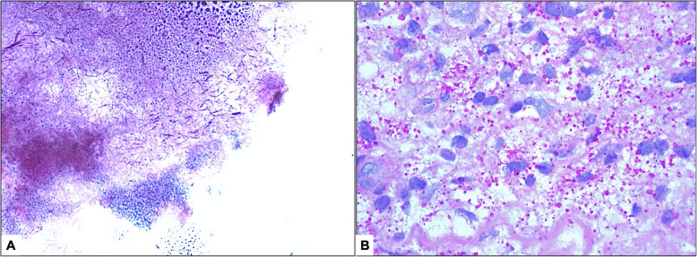
Background: Whipple's disease (WD) is a rare, chronic, infection caused by gram-positive filamentous aerobic actinobacterium Tropheryma whipplei occurs classically in the gastrointestinal tract and shows histopathologically foamy macrophages with typical numerous PAS-positive, non-acid fast particles. Ocular WD in the form of uveitis may occur in the absence of systemic disease but has not been reported to present with scleral manifestation. We describe for the first time to the best of our knowledge 2 cases of scleral nodules with typical histopathological morphology of WD and without systemic involvement.
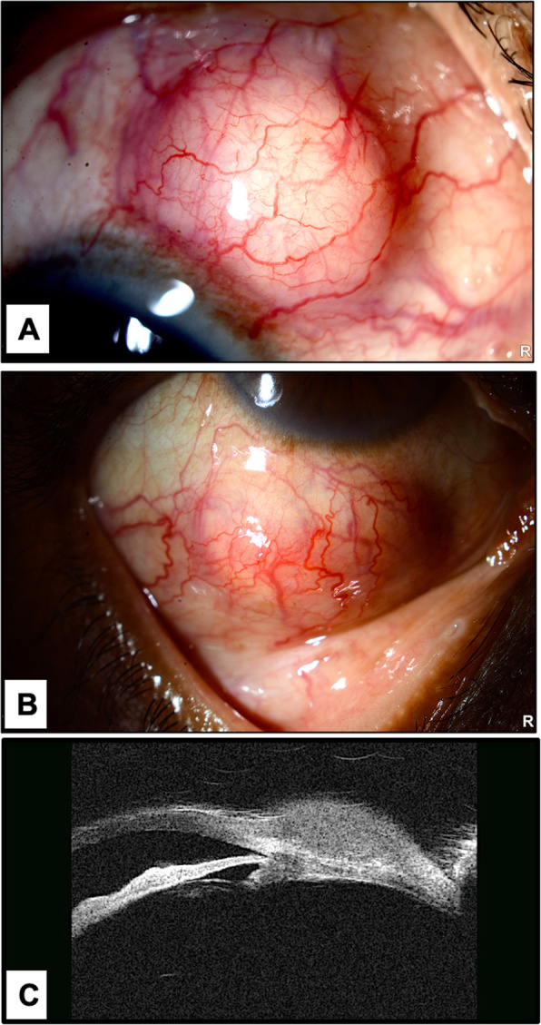
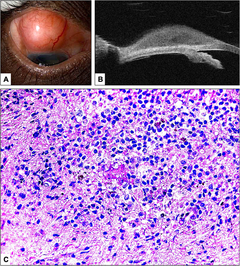
Case presentation: The first was a 53-year old diabetic male farmer who presented with 2 nontender right eye scleral nodules for 3 months, had a negative systemic workup, and surgical excision showed Periodic acid Schiff (PAS)-positive eosinophilic structures inside macrophages. Grocott's methenamine silver (GMS) stain and acid-fast bacilli (AFB) stain of the tissue itself were negative. The second case was a 60-year old male who presented with an asymptomatic superior scleral nodule for 4 months, which showed similar appearance and negative GMS and AFB stains.
Conclusion: WD should be included in the differential diagnosis of scleral nodules even in the absence of systemic symptoms. Surgical excision without systemic treatment resulted in successful outcome without recurrence.
Keywords: Scleral nodule; Scleritis; Tropheryma whipplei; Whipple’s disease.
Conflict of interest statement
Authors declare that there are no competing interests related to this work.
Figures
Similar articles
-
Eyelid abscess as an initial manifestation of whipple's disease: a case report and comprehensive literature review.BMC Ophthalmol. 2025 Mar 6;25(1):111. doi: 10.1186/s12886-025-03902-6. BMC Ophthalmol. 2025. PMID: 40050769 Free PMC article. Review.
-
Ocular Whipple Disease: Cases Diagnosed Over Four Decades.Ocul Immunol Inflamm. 2024 Oct;32(8):1863-1868. doi: 10.1080/09273948.2023.2271995. Epub 2023 Nov 2. Ocul Immunol Inflamm. 2024. PMID: 37917881
-
Whipple's arthritis.Joint Bone Spine. 2016 Dec;83(6):631-635. doi: 10.1016/j.jbspin.2016.07.001. Epub 2016 Aug 5. Joint Bone Spine. 2016. PMID: 27502365 Review.
-
[Whipple's disease and Tropheryma whipplei infections in internal medicine. When to think about it? How to treat?].Rev Med Interne. 2014 Dec;35(12):801-7. doi: 10.1016/j.revmed.2014.04.016. Epub 2014 Jun 2. Rev Med Interne. 2014. PMID: 24933290 Review. French.
-
Whipple's in the valleys: a case of Whipple's with thrombocytopenia and endocarditis.J Clin Pathol. 2014 May;67(5):445-8. doi: 10.1136/jclinpath-2013-201915. Epub 2014 Jan 23. J Clin Pathol. 2014. PMID: 24459171
Cited by
-
Immune recovery uveitis in Whipple's disease: an unusual ocular presentation.J Ophthalmic Inflamm Infect. 2024 Feb 13;14(1):10. doi: 10.1186/s12348-024-00390-5. J Ophthalmic Inflamm Infect. 2024. PMID: 38347376 Free PMC article.
-
Eyelid abscess as an initial manifestation of whipple's disease: a case report and comprehensive literature review.BMC Ophthalmol. 2025 Mar 6;25(1):111. doi: 10.1186/s12886-025-03902-6. BMC Ophthalmol. 2025. PMID: 40050769 Free PMC article. Review.
References
Publication types
MeSH terms
LinkOut - more resources
Full Text Sources
