Review
. 2020 Oct;122(7):151622.
doi: 10.1016/j.acthis.2020.151622. Epub 2020 Sep 9.

Marking vertebrates langerhans cells, from fish to mammals

Affiliations
Review

Marking vertebrates langerhans cells, from fish to mammals

Alessio Alesci et al. Acta Histochem. 2020 Oct.

Abstract

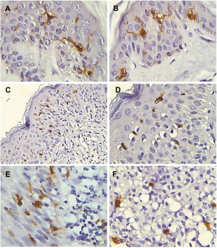
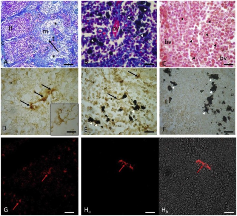
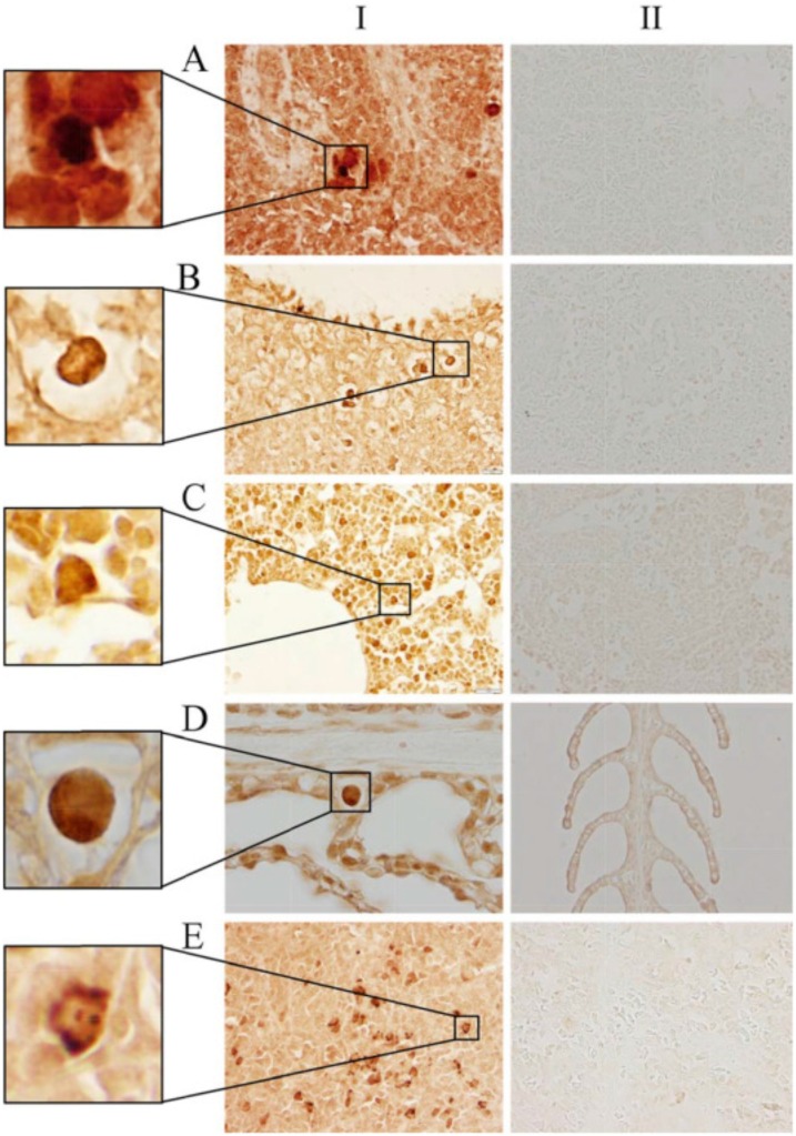
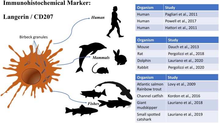
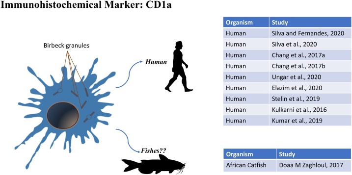
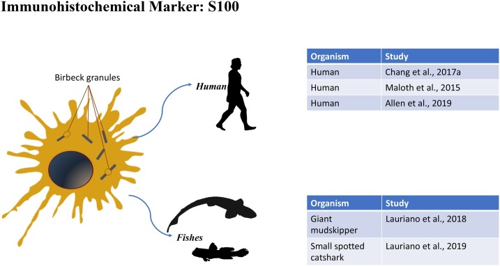
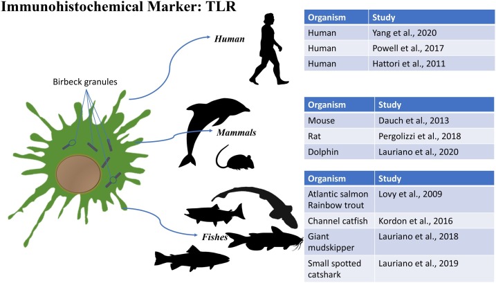
Langerhans cells (LCs) are specialized dendritic cells (DCs) that play a defense role in recognizing foreign antigens, in tissue where antigenic exposures occur, as in the skin and mucous membranes. LCs are able to continuously move within the tissues thanks to dendritic contraction and distension performing their surveillance and/or phagocytosis role. These cells are characterized by the presence of Birbeck granules in their cytoplasm, involved in endocytosis. LCs have been characterized in several classes of vertebrates, from fish to mammals using different histological and molecular techniques. The aim of the present review is to define the state of art and the need of information about immunohistochemical markers of LCs in different classes of vertebrates. The most used immunohistochemical (IHC) markers are Langerin/CD207, CD1a, S-100 and TLR. These IHC markers are described in relation to their finding in different vertebrate classes with phylogenetical considerations. Among the four markers, Langerin/CD207 and TLR have the widest spectrum of cross reactivity in LCs.

Keywords: Dendritic cells; Fish; Immunohistochemistry; Mammals; Markers.


Conflict of interest statement

The authors have no conflict of interest to declare.

Figures

Fig. 1
Fig. 1
Immunohistochemical detection of langerin positive in Human normal skin and in cutaneous lesions. Positive langerin cells with long and short dendrites in the epidermis of normal skin (A, B); langerin positive cells with short dendrites slightly distributed in PCM skin lesions (C, D); positive Langerin cells distributed in the inflammatory infiltrate in the epidermis / dermis interface (E) and dermis (F). Streptavidin-biotin peroxidase method - x200 (C) and x400 (A, B, D, E, F). Reproduction from Pagliari et al., 2011. ELSEVIER LICENSE N° 4881280962191.
Fig. 2
Fig. 2
Immunoperoxidase and confocal immunofluorescence reactions with Langerin/DC 207 in dolphin (Stenella coeruleoalba) lymph nodes. The lymph node parenchyma capsule (c); the projections of the collagen septa that infiltrate the central portion of the lymph nodes dividing the parenchyma into the lymphoid follicles (If) in the cortex; the medulla (m) shows few immune cells (*) separated by sinusoids (black arrow) and trabeculae (t). Mallory's trichome stain (A). Alveolar macrophages (white arrowheads) and lymphocytic cords (white asterisks) are evident in the lymphoid follicles. Mallory's trichome stain (B). Blood vessels (bv) and granular eosinophils (black arrowheads). Hematoxylin/Eosin staining (C). Langerin/CD207 positive dendritic cells (black arrows and insert). Immunoperoxidase method (D). Langerin/CD207 positive dendritic cells (black arrow) in close contact with alveolar macrophages (white arrowhead). Immunoperoxidase method (E). Langerin/CD207 positive dendritic cells (red arrows) in the cortical region of the lymph nodes. Immunofluorescence method (G-H). Control experiments were conducted excluding the primary antibody (F). Magnifications: A 20x; B-C-d-E-F 100x, G–H 40 × . Scale bars: 3A (50 μm), 3B-C-d-E-F-G-H (20 μm). Reproduction from Lauriano et al., 2020. ELSEVIER LICENSE N° 4881300568867 (For interpretation of the references to colour in this figure legend, the reader is referred to the web version of this article).
Fig. 3
Fig. 3
Langerin/CD207 positive cells in the spleen, anterior kidney, and gill of catfish and in Rainbow trout spleen with pAbs in different concentrations; the inserts on the left are magnified images of selected areas. Panel I: Catfish spleen, Abs dilution 1:50 (A); Catfish spleen, Abs dilution 1: 500 (B); Catfish anterior kidney, Abs dilution 1: 500 (C); Catfish gill, Abs dilution 1: 500 (D); Rainbow trout spleen (positive control), Abs dilution 1:50 (E). Panel II shows the respective negative controls (normal goat IgG). Immunoperoxidase method. Photomicrographs (600x). Reproduction from Kordon et al., 2016. ELSEVIER LICENSE N° 4881300043294.
Fig. 4
Fig. 4
Schematic representation of Langerin CD/207 cross reactivity on different vertebrate classes DCs.
Fig. 5
Fig. 5
Schematic representation of CD1a cross reactivity on vertebrate classes DCs.
Fig. 6
Fig. 6
Schematic representation of S100 cross reactivity on different vertebrate classes DCs.
Fig. 7
Fig. 7
Schematic representation of TLR cross reactivity on different vertebrate classes DCs.

References

    1. Abd Elazim N.E., Makboul R., Botros S.N., Awad S.M. Cryopeeling versus trichloroacetic acid peeling in the treatment of solar lentigines: effect on epidermal Langerhans cells. Dermatol. Ther. 2020;33 doi: 10.1111/dth.13288. - DOI - PubMed
    1. Alberts B., Johnson A., Lewis J., et al. Garland Science;; New York: 2002. Molecular Biology of the Cell. 4th edition. Innate Immunity.
    1. Alesci A., Cicero N., Salvo A., Palombieri D., Zaccone D., Dugo G., Bruno M., Vadalà R., Lauriano E.R., Pergolizzi S. Extracts deriving from olive mill waste water and their effects on the liver of the goldfish Carassius auratus fed with hypercholesterolemic diet. Nat. Prod. Res. 2014;28:1343–1349. doi: 10.1080/14786419.2014.903479. - DOI - PubMed
    1. Allen A., Matrova E., Ozgen B., Redleaf M., Emmadi R., Saran N. Langerhans’ cell histiocytosis of the temporal bone in an adult with central diabetes insipidus. Radiol. Case Reports. 2019;14:847–850. doi: 10.1016/j.radcr.2019.03.040. - DOI - PMC - PubMed
    1. Baker M. Antibody anarchy: a call to order. Nature. 2015;527:545–551. doi: 10.1038/527545a. - DOI - PubMed

LinkOut - more resources