

Malignant Cerebral Ischemia in A COVID-19 Infected Patient: Case Review and Histopathological Findings
- PMID: 33066910
- PMCID: PMC7405863
- DOI: 10.1016/j.jstrokecerebrovasdis.2020.105231
Malignant Cerebral Ischemia in A COVID-19 Infected Patient: Case Review and Histopathological Findings
Abstract
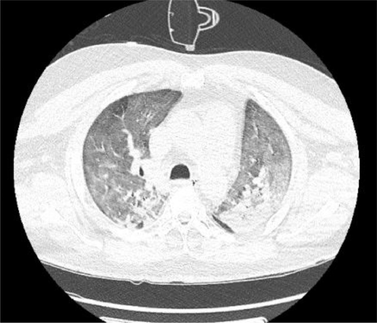
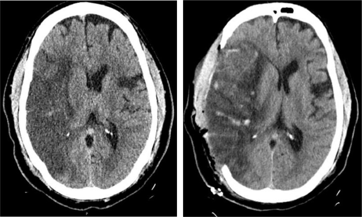
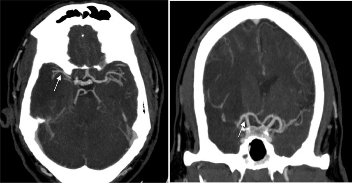
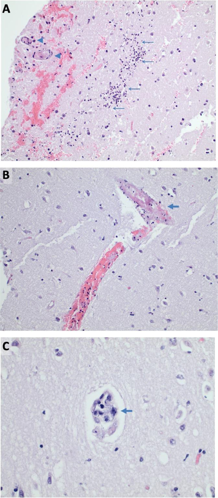
Severe acute respiratory syndrome coronavirus (SARS-CoV-2) is responsible for an unprecedented worldwide pandemic that has severely impacted the United States. As the pandemic continues, a growing body of evidence suggests that infected patients may develop significant coagulopathy with resultant thromboembolic complications including deep vein thrombosis, pulmonary embolism, myocardial infarction, and ischemic stroke. However, this data is limited and comes from recent small case series and observational studies on stroke types, mechanisms, and outcomes.1-14 Furthermore, evidence on the role of therapeutic anticoagulation in SARS-CoV-2 infected patients with elevated inflammatory markers, such as D-dimer, is also limited. We report the case of a middle-aged patient who presented with a large vessel ischemic stroke likely resulting from an underlying inflammatory response in the setting of known novel coronavirus infection (COVID-19). Histopathologic analysis of the patient's ischemic brain tissue revealed hypoxic neurons, significant edema from the underlying ischemic insult, fibrin thrombi in small vessels, and fibroid necrosis of the vascular wall without any signs of vasculature inflammation. Brain biopsy was negative for the presence of SARS-CoV-2 RNA (RT-PCR assay). Along with a growing body of literature, our case suggests that cerebrovascular thromboembolic events in COVID-19 infection may be related to acquired hypercoagulability and coagulation cascade activation due to the release of inflammatory markers and cytokines, rather than virus-induced vasculitis. Further studies to investigate the mechanism of cerebrovascular thromboembolic events and their prevention is warranted.
Keywords: COVID-19; Corona virus; Inflammatory conditions; Ischemic stroke; SARS-CoV-2 RNA; anticoagulation; cerebral sinus thrombosis; cerebrovascular disease; hemorrhagic stroke; thromboembolic conditions; thrombotic conditions; vasculitis.
Copyright © 2020 Elsevier Inc. All rights reserved.
Figures
Similar articles
-
COVID-19 and acute ischemic stroke - A case series from Dubai, UAE.Int J Stroke. 2020 Aug;15(6):699-700. doi: 10.1177/1747493020938285. Epub 2020 Jun 26. Int J Stroke. 2020. PMID: 32525467 No abstract available.
-
SARS-CoV-2 and Stroke in a New York Healthcare System.Stroke. 2020 Jul;51(7):2002-2011. doi: 10.1161/STROKEAHA.120.030335. Epub 2020 May 20. Stroke. 2020. PMID: 32432996 Free PMC article.
-
Stroke and COVID19: Not only a large-vessel disease.J Stroke Cerebrovasc Dis. 2020 Oct;29(10):105074. doi: 10.1016/j.jstrokecerebrovasdis.2020.105074. Epub 2020 Jun 19. J Stroke Cerebrovasc Dis. 2020. PMID: 32912559 Free PMC article.
-
Inflammation and thrombosis in patients with COVID-19: A prothrombotic and inflammatory disease caused by SARS coronavirus-2.Anatol J Cardiol. 2020 Oct;24(4):224-234. doi: 10.14744/AnatolJCardiol.2020.56727. Anatol J Cardiol. 2020. PMID: 33001051 Free PMC article. Review.
-
Guillain-Barré-Strohl syndrome and COVID-19: Case report and literature review.Neuromuscul Disord. 2020 Oct;30(10):859-861. doi: 10.1016/j.nmd.2020.08.354. Epub 2020 Aug 12. Neuromuscul Disord. 2020. PMID: 32912716 Free PMC article. Review.
Cited by
-
Effectiveness of remote ischaemic conditioning is not affected by hyper-inflammation in a rat model of stroke.Sci Rep. 2024 Sep 5;14(1):20750. doi: 10.1038/s41598-024-71328-z. Sci Rep. 2024. PMID: 39237655 Free PMC article.
-
A case-based systematic review on the SARS-COVID-2-associated cerebrovascular diseases and the possible virus routes of entry.J Neurovirol. 2021 Oct;27(5):691-701. doi: 10.1007/s13365-021-01013-8. Epub 2021 Sep 21. J Neurovirol. 2021. PMID: 34546547 Free PMC article.
-
APOE genotype confers context-dependent neurovascular vulnerability in immune-vascularized human forebrain organoids.bioRxiv [Preprint]. 2025 May 9:2025.05.08.652864. doi: 10.1101/2025.05.08.652864. bioRxiv. 2025. PMID: 40654744 Free PMC article. Preprint.
-
Decompressive hemicraniectomy for acute ischemic stroke associated with coronavirus disease 2019 infection: Case report and systematic review.Surg Neurol Int. 2021 Mar 24;12:116. doi: 10.25259/SNI_64_2021. eCollection 2021. Surg Neurol Int. 2021. PMID: 33880221 Free PMC article.
-
Age-Associated Neurological Complications of COVID-19: A Systematic Review and Meta-Analysis.Front Aging Neurosci. 2021 Aug 2;13:653694. doi: 10.3389/fnagi.2021.653694. eCollection 2021. Front Aging Neurosci. 2021. PMID: 34408638 Free PMC article.
References
Publication types
MeSH terms
LinkOut - more resources
Full Text Sources
Medical
Research Materials
Miscellaneous
