. 2020 Oct 16;6(42):eabc5630.
doi: 10.1126/sciadv.abc5630. Print 2020 Oct.

Electrothermal soft manipulator enabling safe transport and handling of thin cell/tissue sheets and bioelectronic devices

Affiliations

Electrothermal soft manipulator enabling safe transport and handling of thin cell/tissue sheets and bioelectronic devices

Byoung Soo Kim et al. Sci Adv. .

Abstract

"Living" cell sheets or bioelectronic chips have great potentials to improve the quality of diagnostics and therapies. However, handling these thin and delicate materials remains a grand challenge because the external force applied for gripping and releasing can easily deform or damage the materials. This study presents a soft manipulator that can manipulate and transport cell/tissue sheets and ultrathin wearable biosensing devices seamlessly by recapitulating how a cephalopod's suction cup works. The soft manipulator consists of an ultrafast thermo-responsive, microchanneled hydrogel layer with tissue-like softness and an electric heater layer. The electric current to the manipulator drives microchannels of the gel to shrink/expand and results in a pressure change through the microchannels. The manipulator can lift/detach an object within 10 s and can be used repeatedly over 50 times. This soft manipulator would be highly useful for safe and reliable assembly and implantation of therapeutic cell/tissue sheets and biosensing devices.


Figures

Fig. 1
Fig. 1. Design of the electrothermal soft manipulator for delicate material transport.
Schematic illustration of (A) the soft, electrothermally controlled manipulator and (B) the process to transport a thin material using the soft manipulator. (A) The soft manipulator consists of a supporter, flexible heater that can convert electrical current to heat, cyanoacrylate-based wet adhesive, and a thermo-responsive PNIPAAm hydrogel with aligned microchannels. (B) Process to transport materials of interests using the soft manipulator. First, the soft manipulator is lowered to let the gel contact a thin material such as a therapeutic cell sheet or an ultrathin film device. During this step, the heater is turned on to contract microchannels of the gel. Second, the heater is turned off to open microchannels of the gel and generate negative pressure in microchannels. As a consequence, the gel serves to hold, lift up, and transport the thin material. Third, the heater is turned on to close microchannels of the gel and, in turn, generate positive pressure in the microchannels. The positive pressure serves to release the thin material onto the target surface.
Fig. 2
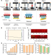
Fig. 2. Fabrication and analysis of rapid temperature-responsive gel.
(A) Schematic illustrating the fabrication process of the gel with anisotropically aligned microchannels. The gel is prepared by directional crystallization and subsequent polymerization. (B) Photograph of the resulting microchanneled hydrogel after swelling in water. (C) Microstructure of the gel: (C-1) scanning electron microscopy (SEM) micrograph of the top surface, (C-2) 3D imaging of the microchanneled hydrogel via micro–computed tomography (micro-CT), and (C-3) SEM micrograph of microchannels that connect the top and bottom of the gel. (D) ESR of gels at different temperatures. (E) Compressive elastic moduli of gels. Samples were compressed in parallel with microchannel direction (axial compression) and perpendicular to microchannel direction (radial compression). (F) Time-dependent volumetric changes of microchanneled gel on heating (F-1) and cooling (F-2). The samples were placed on 40° or 25°C plate. The resulting volumetric change was recorded. (G) Effective diffusion coefficient of water in gels quantified by the reswelling plot (F-2). * represents the statistical significance of the difference of values between conditions indicated with line (*P < 0.01). Photo credit: Byoungsoo Kim, University of Illinois at Urbana-Champaign.
Fig. 3
Fig. 3. Design of soft, electrothermal soft manipulator.
(A) Photograph (top) and a thermal image of the flexible heater captured using an infrared camera (bottom). (B) Temperature change over time at differently applied voltages. The temperature profiles of the heater were obtained using an infrared camera. (C) Structural configuration of the soft manipulator (left) and a photograph of the soft manipulator (right). (D and E) Top: Snapshots of the microchanneled gel in the soft manipulator when the heater was turned on (D) and off (E). Images on the second row represent optical microscopic images of the gel surface when the heater was turned on and off. When the heater was turned on, aligned microchannels of the gel pushed water out while being closed for 20 s (D). When the heater was switched off, the gel in the soft manipulator opened microchannels and pulled water back into microchannels within 20 s (E). Scale bar, 100 μm. Photo credit: Byoungsoo Kim, University of Illinois at Urbana-Champaign.
Fig. 4
Fig. 4. Working mechanism and characterization of the soft manipulator.
(A) Snapshots showing the transport of a 4-inch-diameter silicon wafer using a soft manipulator (upper images). Schematic illustrating the shrinkage and expansion of microchannels and subsequent water movement in microchannels controlled by the electrothermal signal (bottom images). The operating power of the soft manipulator was 5 W. (B) The time-dependent variation of normal adhesion strength measured by the dynamic mechanical analyzer (DMA) during stages 2 and 3 in (A). An initial contact strength of 0.05 kPa was applied to the soft manipulator for this measurement. (C) Fluorescence images of water in microchannels of the gel. The image was obtained from a 3D z-stack confocal microscope before (top) and after adhesion (bottom) of the soft manipulator to a target surface. The heater was attached to the upper part of the gel. (D) Dependency of the adhesion strengths on the initial load. (E) Variation in the adhesion strength as a function of cycle number. (F) Adhesion strength of the soft manipulator measured with the various target substrates in water and air. An initial contact strength of 0.5 kPa was applied to the soft manipulator using DMA for this measurement. Photo credit: Byoungsoo Kim, University of Illinois at Urbana-Champaign.
Fig. 5
Fig. 5. Demonstration of the ability of the soft manipulator to transport cell sheets to target sites.
(A) Snapshots of a process to pick up a skeletal myoblast sheet with forceps. The cell sheet was deformed when picking up the sheet using forceps (right). The cell sheet was stained with methylene blue for visualization. (B) Snapshot of a process to transport the skeletal myoblast sheet onto a glass surface using the soft manipulator. (C) Spatial light interference microscopy (SLIM) images of the cell sheet before (left) and after (right) the transfer, showing off-axis diffraction of the cell sheet. (D) Fluorescence image of a multilayered cell sheet consisting of three different myoblast sheets. The multilayered sheet was prepared by stacking cell sheets using the soft manipulator. (E) Snapshots of a process to transport a skeletal myoblast sheet onto a muscle tissue. It took 30 s for the entire transfer process. (F) Photographs of a rat eye before and after transplantation of a stem cell sheet. The cell sheet transplanted to the corneal epithelium of a rat eye using the soft manipulator. It took 30 s for the entire transfer process. (G) Histological examination of the rat eye before (left) and after (right) a stem cell sheet transfer. Hematoxylin and eosin staining revealed that the stem cell sheet was able to be successfully transplanted onto the anterior corneal surface without substantial interface space generation. Photo credit: Byoungsoo Kim, University of Illinois at Urbana-Champaign.
Fig. 6
Fig. 6. Transportation of an ultrathin EP sensor.
(A) Device configuration of the ultrathin EP sensor (t = 1 μm) tailored for the measurement of ECG signals. (B) Snapshot of a process to transport the device to the surface of the pig heart. It took 30 s to capture and deliver the device onto the pig heart. (C) Photograph of the device transplanted to the pig heart using the soft manipulator. (D) Representative ECG signals measured using the transplanted device. Photo credit: Byoungsoo Kim, University of Illinois at Urbana-Champaign.

References

    1. Yang J., Yamato M., Kohno C., Nishimoto A., Sekine H., Fukai F., Okano T., Cell sheet engineering: Recreating tissues without biodegradable scaffolds. Biomaterials 26, 6415–6422 (2005). - PubMed
    1. Yang J., Yamato M., Shimizu T., Sekine H., Ohashi K., Kanzaki M., Ohki T., Nishida K., Okano T., Reconstruction of functional tissues with cell sheet engineering. Biomaterials 28, 5033–5043 (2007). - PubMed
    1. Nishida K., Yamato M., Hayashida Y., Watanabe K., Maeda N., Watanabe H., Yamamoto K., Nagai S., Kikuchi A., Tano Y., Okano T., Functional bioengineered corneal epithellial sheet grafts from corneal stem cells expanded ex vivo on a temperature-responsive cell culture surface. Transplantation 77, 379–385 (2004). - PubMed
    1. Sekine H., Shimizu T., Sakaguchi K., Dobashi I., Wada M., Yamato M., Kobayashi E., Umezu M., Okano T., In vitro fabrication of functional three-dimensional tissues with perfusable blood vessels. Nat. Commun. 4, 1399 (2013). - PMC - PubMed
    1. Kim D.-H., Lu N., Ma R., Kim Y.-S., Kim R.-H., Wang S., Wu J., Won S. M., Tao H., Islam A., Yu K. J., Kim T.-i., Chowdhury R., Ying M., Xu L., Li M., Chung H.-J., Keum H., Cormick M. M., Liu P., Zhang Y.-W., Omenetto F. G., Huang Y., Coleman T., Rogers J. A., Epidermal electronics. Science 333, 838–843 (2011). - PubMed

Publication types

LinkOut - more resources