. 2020 Nov 3;117(44):27329-27338.
doi: 10.1073/pnas.2009206117. Epub 2020 Oct 16.

Mapping glycan-mediated galectin-3 interactions by live cell proximity labeling

Affiliations

Mapping glycan-mediated galectin-3 interactions by live cell proximity labeling

Eugene Joeh et al. Proc Natl Acad Sci U S A. .

Abstract

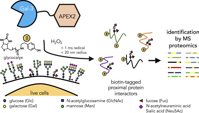
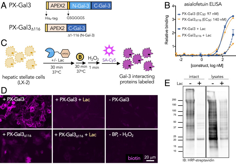
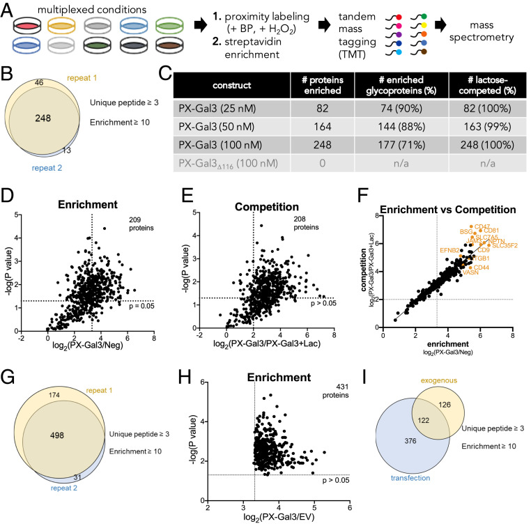
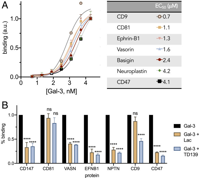
Galectin-3 is a glycan-binding protein (GBP) that binds β-galactoside glycan structures to orchestrate a variety of important biological events, including the activation of hepatic stellate cells and regulation of immune responses. While the requisite glycan epitopes needed to bind galectin-3 have long been elucidated, the cellular glycoproteins that bear these glycan signatures remain unknown. Given the importance of the three-dimensional (3D) arrangement of glycans in dictating GBP interactions, strategies that allow the identification of GBP receptors in live cells, where the native glycan presentation and glycoprotein expression are preserved, have significant advantages over static and artificial systems. Here we describe the integration of a proximity labeling method and quantitative mass spectrometry to map the glycan and glycoprotein interactors for galectin-3 in live human hepatic stellate cells and peripheral blood mononuclear cells. Understanding the identity of the glycoproteins and defining the structures of the glycans will empower efforts to design and develop selective therapeutics to mitigate galectin-3-mediated biological events.

Keywords: galectins; glycan; glycomics; proteomics; proximity labeling.


Conflict of interest statement

The authors declare no competing interest.

Figures

Fig. 1.
Fig. 1.
Schematic illustration of the identification of galectin-3 (Gal-3) interacting proteins by in situ proximity labeling. Recombinant APEX2 and galectin-3 fusion proteins are applied to living cells where galectin-3 can freely diffuse to bind its cognate ligands. On addition of biotin phenol (yellow circle with “B”; 30 min) and hydrogen peroxide (H2O2; 1 min), APEX2 catalyzes the formation of highly-reactive biotin-phenoxyl radicals that react within a short range (<20 nm) of the galectin-3 complex within a short time frame (<1 ms). The biotin-tagged protein interactors can then be identified using MS-based proteomics.
Fig. 2.
Fig. 2.
Design and characterization of PX-Gal3 fusion constructs for proximity labeling in LX-2 HSCs. (A) APEX2 fusion constructs of galectin-3, PX-Gal3 and PX-Gal3Δ116, were constructed. Both proteins include an N-terminal His-tag sequence, followed by the APEX2 enzyme, and either full-length galectin-3 or the C-terminal domain of galectin-3. PX-Gal3Δ116 lacks the N-terminal domain of galectin-3, which was previously implicated for its homo-oligomerization on binding of glycans at the cell surface. (B) Using an ELISA with asialofetuin as a model glycan-bearing ligand, the fusion constructs retained similar glycan-binding activities, as determined by EC50 values. (C) Application of the proximal labeling method to live adherent HSCs. The recombinant fusion proteins were first incubated with HSCs (30 min, 37 °C). After washing to remove unbound proteins, biotin phenol (yellow circle with a B; 500 μM, 30 min, 37 °C) was added, followed by H2O2 (1 mM, 1 min, RT). Biotinylated interactors (purple) were subsequently probed using Cy5-labeled streptavidin. Conditions were optimized according to reported radical-mediated biotinylation procedures (20, 21). (D) Fluorescence microscopy images of biotin-tagged (purple) HSCs showing that PX-Gal3 (100 nM; 30 min) generates significant labeling over negative controls in which a component (e.g., protein or biotin phenol and H2O2) of the labeling protocol is omitted. Coincubation with lactose (Lac; 100 mM) causes a loss of signal, suggesting that the majority of labeled interactions are glycan-dependent. PX-Gal3Δ116 fails to significantly label interactors. (E) Western blotting of (10 μg lysate per lane) biotinylated proteins produced from the proximity labeling method applied to intact live cells vs. cell lysates showing that only a subset of interactors is captured (SI Appendix, Fig. S6).
Fig. 3.
Fig. 3.
Identification and analysis of the PX-Gal3 interactome in LX-2 HSCs by quantitative MS-based proteomics. (A) Experimental workflow for the preparation of samples for analysis from LX-2 HSCs. (B) A total of 248 proteins were enriched across two replicates, defined as proteins with three or more unique peptides and with a TMT ratio (PX-Gal3/Neg) ≥10. Neg indicates conditions in which the cells were not incubated with PX-Gal3 but were still treated with biotin phenol and H2O2. (C) Analysis of proteins enriched by PX-Gal3 by dosage, glycosylation status (assigned by UniProt), and competition with lactose (100 mM). (D) Statistically significant (P < 0.05) and enriched proteins found in cells treated with PX-Gal3 (100 nM, 30 min). (E) Proteins that were statistically significant (P < 0.05) and competed (TMT ratio of PX-Gal3/PX-Gal3+Lac ≥4) on coincubation with exogenous lactose (100 mM). (F) There is high linear correlation between proteins that were enriched by and those that were competed with lactose. (G) A total of 498 proteins were found to be enriched across two replicates when PX-Gal3 was transiently overexpressed in HSCs. (H) A total of 431 proteins were found to be significantly enriched (P < 0.05). (I) There were 122 overlapping proteins between the exogenous and transfection protocols (SI Appendix, Table S1).
Fig. 4.
Fig. 4.
Validation of binding between galectin-3 and proteins identified from proteomics analysis. (A) ELISA binding curves and corresponding EC50 values determined for recombinant proteins against galectin-3. (B) Coincubation of galectin-3 (5 μM) with lactose (Lac, 100 mM) or TD139 (15.4 μM) competes for glycan-mediated binding interactions in ELISA.
Fig. 5.
Fig. 5.
MS-based determination of N-glycan abundance and composition. Composition and relative amounts of the most abundant N-glycans found on LX-2 cell surfaces (black bars), glycoproteins enriched by PX-Gal3 (orange bars), and immunoprecipitated (IP) BSG (blue bars). The most abundant N-glycans found on BSG differ greatly from the cell surface samples. An asterisk indicates N-glycans that were also found in the antibody used for IP. Terminal galactose (yellow circles) presenting N-glycans were found to be enriched in BSG compared with the cell surface. Oligomannose glycans bear terminal mannose residues (green circles) and are designated with an M (M7, M6, M8, M5); all others shown here are complex-type N-glycans.
Fig. 6.
Fig. 6.
Identification and analysis of the PX-Gal3 interactome in PBMCs by quantitative MS-based proteomics. (A) Statistically significant (P < 0.05) and enriched proteins found in cells treated with PX-Gal3 (100 nM, 30 min). (B) Proteins that were statistically significant (P < 0.05) and competed (TMT ratio of PX-Gal3/PX-Gal3+Lac ≥4) on coincubation with exogenous lactose (100 mM). (C) There was high linear correlation between proteins that were enriched by lactose and those that were competed with lactose. (D) A total of 39 proteins were enriched in both LX-2s and PBMCs, whereas 304 proteins were exclusively identified in PBMCs.

References

    1. Di Lella S.et al., When galectins recognize glycans: From biochemistry to physiology and back again. Biochemistry 50, 7842–7857 (2011). - PMC - PubMed
    1. Henderson N. C.et al., Galectin-3 expression and secretion links macrophages to the promotion of renal fibrosis. Am. J. Pathol. 172, 288–298 (2008). - PMC - PubMed
    1. Henderson N. C. M.et al., Galectin-3 regulates myofibroblast activation and hepatic fibrosis. Proc. Natl. Acad. Sci. U.S.A. 103, 5060–5065 (2006). - PMC - PubMed
    1. Rabinovich G. A., Toscano M. A., Turning “sweet” on immunity: Galectin-glycan interactions in immune tolerance and inflammation. Nat. Rev. Immunol. 9, 338–352 (2009). - PubMed
    1. Díaz-Alvarez L., Ortega E., The many roles of galectin-3, a multifaceted molecule, in innate immune responses against pathogens. Mediators Inflamm. 2017, 9247574 (2017). - PMC - PubMed

Publication types