. 2020 Nov 3;117(44):27528-27539.
doi: 10.1073/pnas.2006186117. Epub 2020 Oct 16.

Selective tumor antigen vaccine delivery to human CD169+ antigen-presenting cells using ganglioside-liposomes

Affiliations

Selective tumor antigen vaccine delivery to human CD169+ antigen-presenting cells using ganglioside-liposomes

Alsya J Affandi et al. Proc Natl Acad Sci U S A. .

Abstract

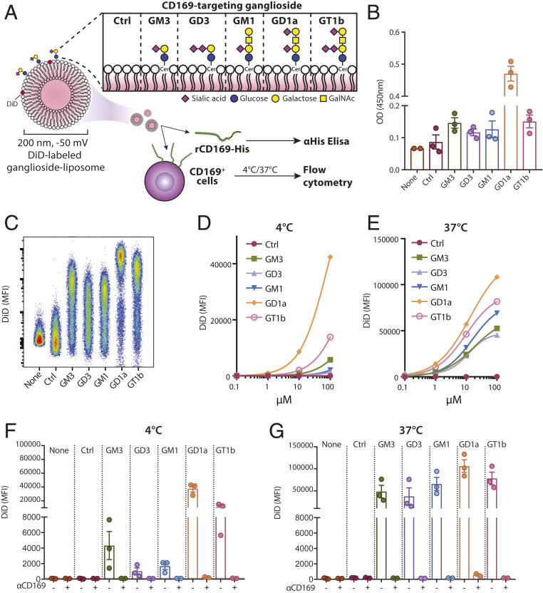
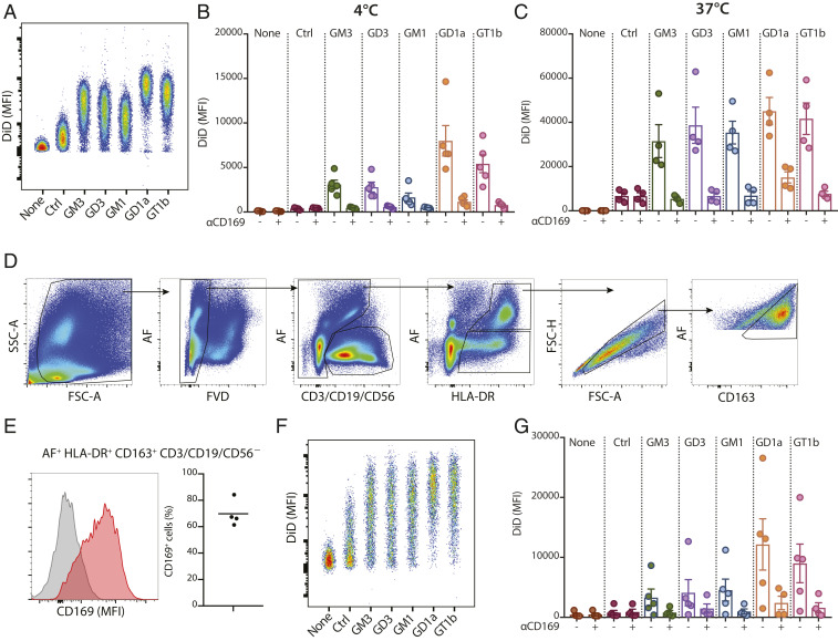
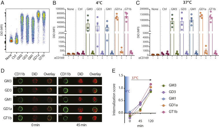
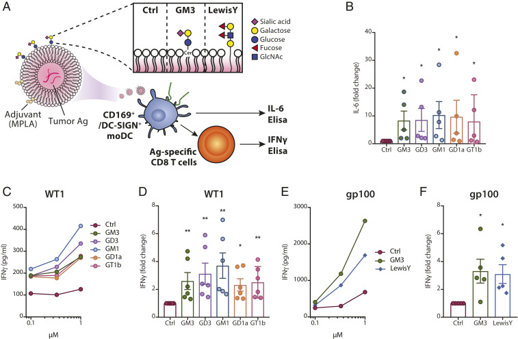
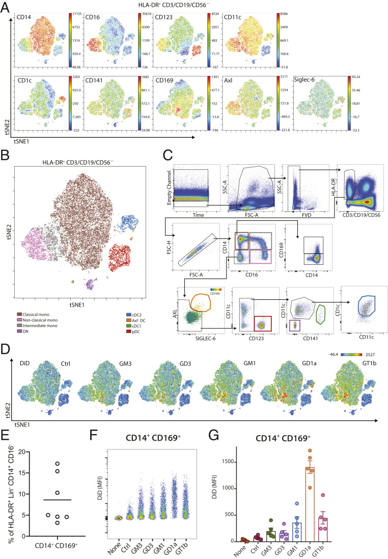
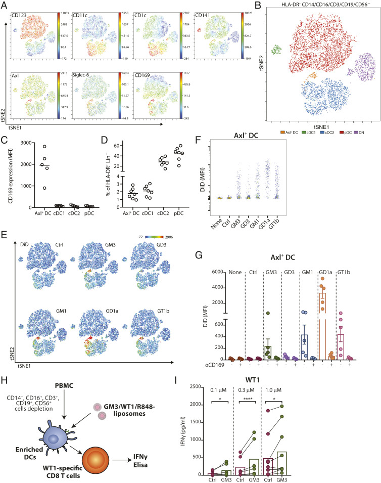
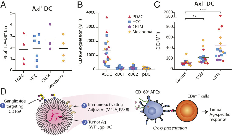
Priming of CD8+ T cells by dendritic cells (DCs) is crucial for the generation of effective antitumor immune responses. Here, we describe a liposomal vaccine carrier that delivers tumor antigens to human CD169/Siglec-1+ antigen-presenting cells using gangliosides as targeting ligands. Ganglioside-liposomes specifically bound to CD169 and were internalized by in vitro-generated monocyte-derived DCs (moDCs) and macrophages and by ex vivo-isolated splenic macrophages in a CD169-dependent manner. In blood, high-dimensional reduction analysis revealed that ganglioside-liposomes specifically targeted CD14+ CD169+ monocytes and Axl+ CD169+ DCs. Liposomal codelivery of tumor antigen and Toll-like receptor ligand to CD169+ moDCs and Axl+ CD169+ DCs led to cytokine production and robust cross-presentation and activation of tumor antigen-specific CD8+ T cells. Finally, Axl+ CD169+ DCs were present in cancer patients and efficiently captured ganglioside-liposomes. Our findings demonstrate a nanovaccine platform targeting CD169+ DCs to drive antitumor T cell responses.

Keywords: CD169; CD8+ T cells; Siglec-1; dendritic cells; vaccination.


Conflict of interest statement

The authors declare no competing interest.

Figures

Fig. 1.
Fig. 1.
Ganglioside-liposomes bind CD169 and CD169-overexpressing THP-1. (A) Gangliosides GM3, GD3, GM1, GD1a, and GT1b were incorporated into liposomes, and binding to CD169 was determined by recombinant CD169 ELISA or cell-based flow cytometry. GalNAc, N-acetyl galactosamine; Cer, ceramide; Ctrl, control. (B) Binding of recombinant human CD169 (rCD169) to ganglioside-liposomes as determined by ELISA. (CG) DiD-labeled ganglioside-liposomes were incubated with THP-1 cells overexpressing CD169 (TSn), and binding at 4 °C or uptake at 37 °C was determined by flow cytometry. (C) Representative plot of ganglioside-liposome binding (100 µM) to TSn. (D and E) Binding or uptake of ganglioside-liposomes at different concentrations from one representative experiment out of two is shown. (F and G) TSn were preincubated with anti-CD169 blocking antibody at 4 °C for 15 min to show specificity of 100 µM ganglioside-liposome binding or uptake. Data are mean ± SEM from three independent experiments.
Fig. 2.
Fig. 2.
Ganglioside-liposomes bind CD169-expressing moMacs and human splenic macrophages. (A and B) DiD-labeled ganglioside-liposomes were incubated with moMacs, and binding (4 °C; A and B) and uptake (37 °C; C) was determined by flow cytometry. Data are mean ± SEM from four independent donors. (D) Human spleen cells were incubated with ganglioside-liposomes at 37 °C for 45 min and stained for cell lineage markers. Gating strategy of autofluorescence+ (AF) HLA-DR+ CD163+ CD3/CD19/CD56 macrophages is displayed. (E) The expression of CD169 on human splenic macrophages is shown as a representative histogram (Left; gray, fluorescence minus one; red, CD169) and percentages (Right; n = 4). (F and G) Ganglioside-liposome uptake by human splenic macrophages as (F) representative dot plot and (G) quantification (n = 4 to 5) is shown. When indicated, macrophages were preincubated with anti-CD169 blocking antibody to block ganglioside-liposome binding. Data are mean ± SEM from n = 4 to 5 donors.
Fig. 3.
Fig. 3.
Ganglioside-liposomes bind to CD169-expressing moDCs. (AC) Ganglioside-liposomes were incubated with IFN-I–treated moDCs, and binding (4 °C; A and B) and uptake (37 °C; C) was determined by flow cytometry. Ctrl, control. In some conditions, moDCs were preincubated with anti-CD169 blocking antibody. Data are mean ± SEM from four donors. (D and E) After 45 min binding (4 °C), ganglioside-liposome internalization at 37 °C was measured by imaging cytometry. (D) Representative images and (E) quantification of internalization are shown. Data are representative of two independent experiments from two donors.
Fig. 4.
Fig. 4.
Adjuvant/tumor antigen-containing ganglioside-liposomes activate CD169-expressing moDCs and are cross-presented to antigen-specific T cells. (A) Ganglioside- (i.e., GM3) or Lewis Y-liposomes with MPLA and/or tumor-associated antigen for moDC activation or antigen presentation assay. GlcNAc, N-acetylglucosamine. (B) moDCs were incubated with MPLA-containing ganglioside-liposomes at 4 °C and washed, and IL-6 secretion after 24 h was measured by ELISA. Values are calculated as fold change over control liposome; data are mean ± SEM from five donors. (C and D) Ganglioside-liposomes containing short WT1 peptide and MPLA were loaded to moDCs and washed away, and WT1-specific CD8+ T cells were added. IFNγ secretion after 24 h was determined by ELISA. (C) Secreted IFNγ at different doses of liposomes are shown (data pooled from n = 6 donors). (D) Production of IFNγ after exposure to 1 µM ganglioside-liposomes normalized to control liposomes. Data are mean ± SEM from six donors. (E and F) GM3- or Lewis Y-liposome containing long gp100 peptide was loaded to moDCs, followed by 24 h coculture with gp100-specific T cell clone. IFNγ production by gp100-specific T cells (E) at different doses of liposomes (pooled from five donors) and (F) after treatment with 1 µM GM3- or Lewis Y-liposome normalized to control liposome. Data are mean ± SEM from five donors. Kruskal–Wallis multiple two-tailed t tests using a two-stage linear step-up procedure of Benjamini, Krieger, and Yekutieli, with Q = 0.05, was used (*adjusted P < 0.05, **adjusted P < 0.01).
Fig. 5.
Fig. 5.
Ganglioside-liposomes target human CD14+ CD169+ monocytes. (A) High-dimensionality reduction analysis of circulating HLA-DR+ CD3/CD19/CD56 cells using the expression of monocytes and DC subset markers CD14, CD16, CD123, CD11c, CD1c, CD141, Axl, Siglec-6, and CD169 using viSNE analysis. (B) Overlay of viSNE map with (C) conventional gating identifies classical monocytes, nonclassical monocytes, intermediate monocytes, plasmacytoid DCs (pDC), conventional DC1 (cDC1), conventional DC2 (cDC2), and Axl+ Siglec-6+ DCs (Axl+ DC). DN defines CD11c CD123 cells. (D) Uptake of DiD-labeled ganglioside-liposomes is restricted to CD169+ populations on viSNE map. Ctrl, control. (E) Percentage of CD14+ CD169+ cells within classical monocytes (HLA-DR+ CD14+ CD16 Lin cells; n = 7). (F and G) Ganglioside-liposome uptake by human CD14+ CD169+ monocytes as (F) representative plot and (G) quantification (n = 5) is shown. Data are mean ± SEM from five donors.
Fig. 6.
Fig. 6.
Human Axl+ DCs take up ganglioside-liposomes and stimulate CD8+ T cells. (A) High-dimensionality reduction analysis of circulating HLA-DR+ CD3/CD19/CD56/CD14/CD16 cells using the expression of DC subsets markers CD123, CD11c, CD1c, CD141, Axl, Siglec-6, and CD169 using viSNE analysis. (B) Overlay of viSNE map with conventional gating identifies plasmacytoid DCs (pDC), conventional DC1 (cDC1), conventional DC2 (cDC2), and Axl+ Siglec-6+ DCs (Axl+ DC). DN defines CD11cCD123 cells. (C) The expression of CD169 on DC subsets is shown (n = 5). (D) Distribution of DC populations within HLA-DR+ Lin(CD3/CD19/CD56/CD14/CD16) cells (n = 7). (E) Uptake of DiD-labeled ganglioside-liposomes is restricted to Axl+ DC population on viSNE map. (F and G) Ganglioside-liposome uptake by human Axl+ DCs as (F) representative plot and (G) quantification (n = 5) is shown. Ctrl, control. In some conditions, cells were preincubated with anti-CD169 blocking antibody to block ganglioside-liposome binding. Data are mean ± SEM from n = 4 to 5 donors. (H and I) After lineage depletion, enriched DCs were incubated with different concentrations of GM3/WT1/R848 liposome or control liposome at 37 °C and washed, and WT1-specific CD8+ T cells were added. (I) IFNγ secretion after 24 h was determined by ELISA. Data are means from six to nine donors (paired t test: *P < 0.05, ****P < 0.001).
Fig. 7.
Fig. 7.
Axl+ DCs are present in cancer patients and can be targeted by ganglioside-liposomes. (A) Percentage of Axl+ DCs within HLA-DR+ Lin(CD3/CD19/CD56/CD14/CD16) cells in patients with pancreatic ductal adenocarcinoma (PDAC; n = 4), hepatocellular carcinoma (HCC; n = 7), colorectal liver metastasis (CRLM; n = 3), and melanoma (n = 4). (B) The expression of CD169 on DC subsets of cancer patients is shown. (C) Ganglioside-liposome uptake by Axl+ DCs of cancer patients is shown. Friedman test using a two-stage linear step-up procedure of Benjamini, Krieger, and Yekutieli, with Q = 0.05, was used (**adjusted P < 0.01, ****adjusted P < 0.0001). (D) In this study, we have designed nanovaccine carriers targeting CD169+ APCs, particularly Axl+ DCs, using liposomes that contain (1) gangliosides to target CD169, (2) immune-activating adjuvant, and (3) encapsulated tumor antigen. Uptake of ganglioside-liposomes by CD169+ DCs will lead to cross-presentation and activation of tumor-specific CD8+ T cells.

References

    1. Haslam A., Prasad V., Estimation of the percentage of US patients with cancer who are eligible for and respond to checkpoint inhibitor immunotherapy drugs. JAMA Netw. Open 2, e192535 (2019). - PMC - PubMed
    1. Yarchoan M., Hopkins A., Jaffee E. M., Tumor mutational burden and response rate to PD-1 inhibition. N. Engl. J. Med. 377, 2500–2501 (2017). - PMC - PubMed
    1. Nevala-Plagemann C., Hidalgo M., Garrido-Laguna I., From state-of-the-art treatments to novel therapies for advanced-stage pancreatic cancer. Nat. Rev. Clin. Oncol. (2019). - PubMed
    1. Wu A. A., Jaffee E., Lee V., Current status of immunotherapies for treating pancreatic cancer. Curr. Oncol. Rep. 21, 60 (2019). - PubMed
    1. Vonderheide R. H., The immune revolution: A case for priming, not checkpoint. Cancer Cell 33, 563–569 (2018). - PMC - PubMed

Publication types

MeSH terms