. 2021 Mar;78(6):2821-2838.
doi: 10.1007/s00018-020-03665-x. Epub 2020 Oct 16.

Wip1 controls the translocation of the chromosomal passenger complex to the central spindle for faithful mitotic exit

Affiliations

Wip1 controls the translocation of the chromosomal passenger complex to the central spindle for faithful mitotic exit

Xianghua Zhang et al. Cell Mol Life Sci. 2021 Mar.

Abstract

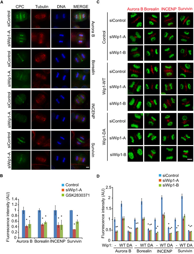
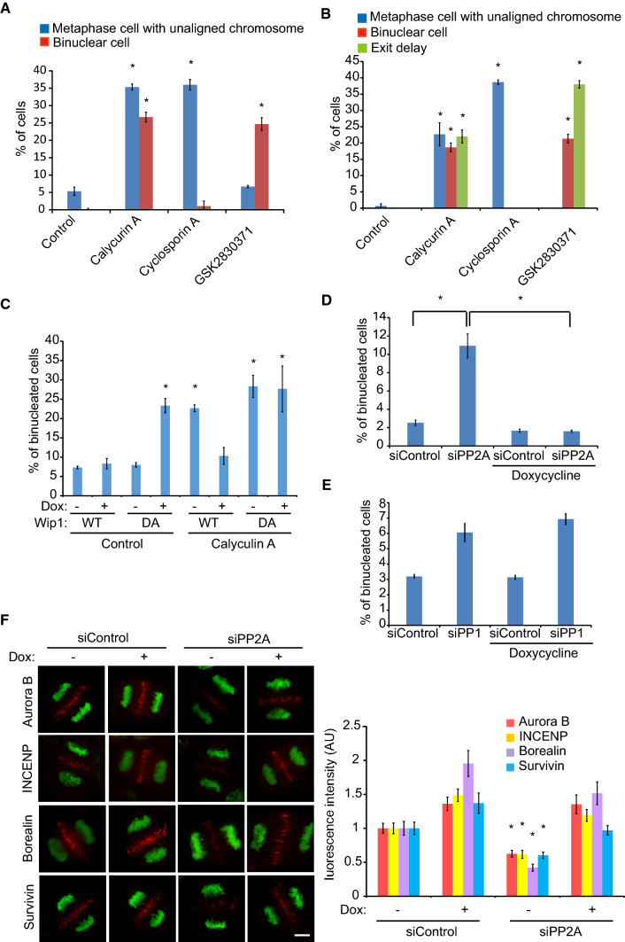
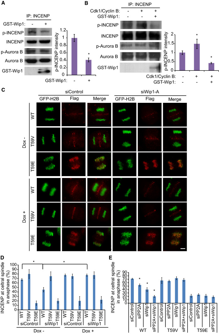
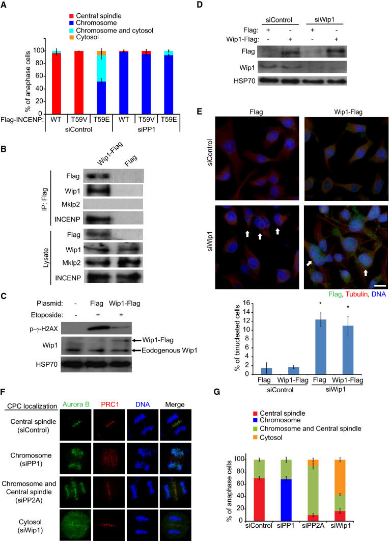
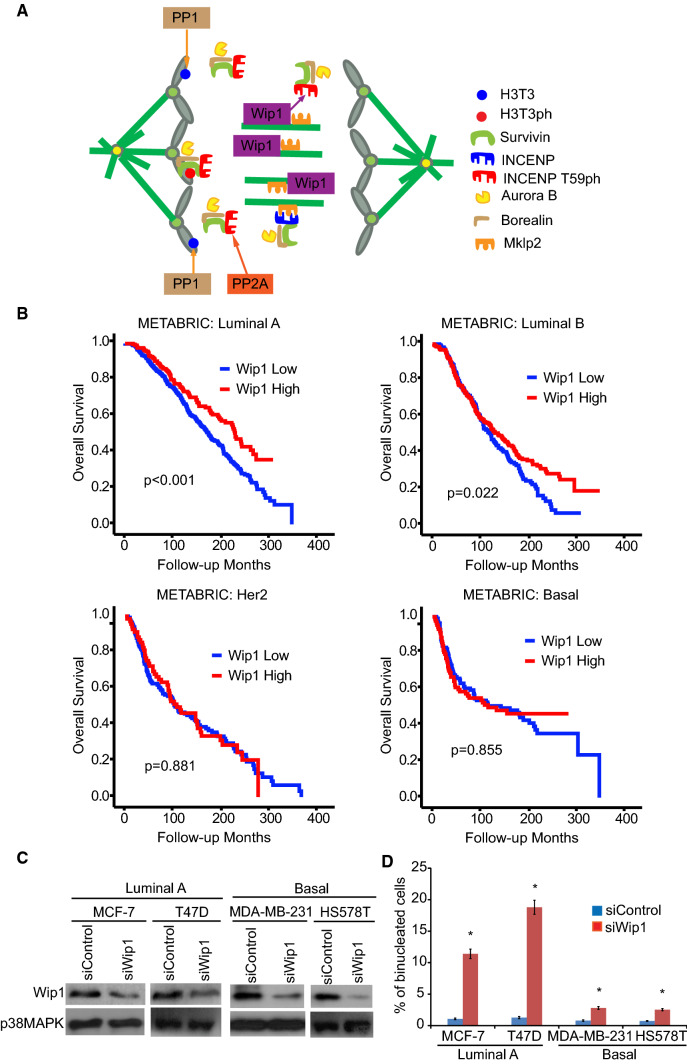
Dramatic cellular reorganization in mitosis critically depends on the timely and temporal phosphorylation of a broad range of proteins, which is mediated by the activation of the mitotic kinases and repression of counteracting phosphatases. The mitosis-to-interphase transition, which is termed mitotic exit, involves the removal of mitotic phosphorylation by protein phosphatases. Although protein phosphatase 1 (PP1) and protein phosphatase 2A (PP2A) drive this reversal in animal cells, the phosphatase network associated with ordered bulk dephosphorylation in mitotic exit is not fully understood. Here, we describe a new mitotic phosphatase relay in which Wip1/PPM1D phosphatase activity is essential for chromosomal passenger complex (CPC) translocation to the anaphase central spindle after release from the chromosome via PP1-mediated dephosphorylation of histone H3T3. Depletion of endogenous Wip1 and overexpression of the phosphatase-dead mutant disturbed CPC translocation to the central spindle, leading to failure of cytokinesis. While Wip1 was degraded in early mitosis, its levels recovered in anaphase and the protein functioned as a Cdk1-counteracting phosphatase at the anaphase central spindle and midbody. Mechanistically, Wip1 dephosphorylated Thr-59 in inner centromere protein (INCENP), which, subsequently bound to MKLP2 and recruited other components to the central spindle. Furthermore, Wip1 overexpression is associated with the overall survival rate of patients with breast cancer, suggesting that Wip1 not only functions as a weak oncogene in the DNA damage network but also as a tumor suppressor in mitotic exit. Altogether, our findings reveal that sequential dephosphorylation of mitotic phosphatases provides spatiotemporal regulation of mitotic exit to prevent tumor initiation and progression.

Keywords: Aurora B; Checkpoint; DNA damage response; Homeostasis; MKLP1.


Conflict of interest statement

The authors declare that they have no conflict of interest.

Figures

Fig. 1
Fig. 1
Wip1 is an essential phosphatase for mitotic exit. a Overall survival curves of patients stratified according to the expression of the Wip1 mRNA in the METABRIC database. b, c HeLa cells were synchronized by a double thymidine block (b) or thymidine-nocodazole block (c), released into fresh media, and harvested at the indicated times. Cell lysates were analyzed via Western blotting with the indicated antibodies. Relative intensities of the bands were measured with image processing software (Image Studio ver5.0). The mitotic index (MI) was determined by fluorescence-activated cell sorting (FACS) with anti-MPM2 antibody staining. Hsp70 served as a loading control. d HeLa cells were fixed with MeOH and stained with the indicated antibodies. Images are maximum projections from z stacks of representative cells stained for Wip1 (green), β-tubulin or Plk1 (red), and DNA (blue). Scale bars, 5 μm. e HeLa cells were transfected with control (siControl) or Wip1-specific siRNAs (siWip1-A and siWip1-B) and harvested at 72 h after transfection. Lysates were analyzed by Western blotting with the indicated antibodies. f, g HeLa/GFP-histone H2B cells were treated with siWip1-A or the Wip1 inhibitor GSK2830371, and imaged for GFP-histone H2B using time-lapse microscopy starting at 60 h after transfection. Images were captured every 3 min to monitor mitotic progression. The duration from nuclear envelope breakdown (NEB) to chromosome segregation (NEB to anaphase onset) and from anaphase onset to chromosome decondensation (mitotic exit) were measured for control and Wip1-depleted cells (n = 40 cells). Still frames from time-lapse movies of representative cells are shown in (g). Scale bar, 10 μm. h, i HeLa/GFP-histone H2B cells stably transfected with tetracycline-inducible WT Wip1 or the DA mutant were transfected with siControl or siWip1. The cells were treated with doxycycline to induce Wip1 protein expression at 48 h post-transfection and fixed with methanol. After staining with a β-tubulin antibody, binucleated cells were counted in three independent experiments (n = 500 interphase cells for each quantification). Scale bar, 20 μm. Error bars, SEM. *p < 0.01 (two-tailed t-test)
Fig. 2
Fig. 2
Wip1 is required for CPC translocation to the central spindle in anaphase. a, b After treatment with siWip1-A for 72 h or the Wip1 inhibitor for 24 h, HeLa cells were stained with antibodies against CPC components, and the intensity was determined from 30 anaphase cells in three independent experiments. c, d Forty-eight hours after siRNA transfection, HeLa/GFP-histone H2B cells stably transfected with tetracycline-inducible WT Wip1 or the DA mutant were treated with doxycycline for 24 h to induce Wip1 protein expression. The cells were stained with a β-tubulin antibody and the intensity of CPC components was determined from 30 anaphase cells in three independent experiments. Scale bars, 5 μm. Error bars, SEM. *p < 0.01 (two-tailed t-test)
Fig. 3
Fig. 3
MKLP2 acts as a docking site for Wip1 at the central spindle in anaphase. a After treatment with siWip1-A or -B for 72 h or the Wip1 inhibitor for 24 h, HeLa cells were stained with the indicated antibodies and the intensity was determined from 30 anaphase cells in three independent experiments. b Lysates of HeLa cells were subjected to IP with an INCENP antibody followed by Western blotting. ce Forty-eight hours after siRNA transfection, cells were prepared as described in Fig. 2c. The lysates were subjected to Western blotting (c). Cells were stained with an Mklp2 (d) or Wip1 antibody (e). The intensities of Mklp2 (d) and Wip1 (e) were determined from ten anaphase cells in three independent experiments. Scale bars, 5 μm. Error bars, SEM. *p < 0.01 (two-tailed t-test)
Fig. 4
Fig. 4
Wip1 compensates for the lack of PP2A during mitotic exit. a HeLa cells were treated with the PP1/PP2A inhibitor calyculin A for 5 h, the PP2B inhibitor cyclosporin A for 5 h, or the Wip1 inhibitor GSK2830371for 24 h. After staining with a β-tubulin antibody, binucleated cells and metaphase cells containing unaligned chromosomes were counted in three independent experiments (n = 500 interphase cells for each quantification). b HeLa/GFP-histone H2B cells were treated with the indicated inhibitors and imaged for GFP-histone H2B by time-lapse microscopy. Images were captured every 3 min to monitor mitotic progression. Metaphase cells containing unaligned chromosomes, binucleated cells, and cytokinetic cells showing delayed mitotic exit (more than 40 min) were counted and plotted. c After inducing WT Wip1 or DA mutant protein, the cells were treated with calyculin A for 5 h and fixed with methanol. After staining with a β-tubulin antibody, binucleated cells were counted in three independent experiments (n = 500 interphase cells for each quantification). df Forty-eight hours after siPP1 or siPP2A transfection, HeLa/GFP-histone H2B cells stably transfected with tetracycline-inducible WT Wip1 were treated with doxycycline for 24 h to induce Wip1 protein expression. After staining with a β-tubulin antibody, binucleated cells were counted in three independent experiments (d, e, n = 500 interphase cells for each quantification). The cells were also stained with antibodies against CPC components, and the intensity was determined from 30 anaphase cells in three independent experiments (f). Scale bar, 5 μm. Error bars, SEM. *p < 0.01 (two-tailed t-test)
Fig. 5
Fig. 5
Wip1 dephosphorylates Thr59 in INCENP for CPC translocation to the central spindle. a Thymidine-nocodazole arrested HeLa cells were treated with calyculin A for 5 h and then released into fresh media for 2 h. The lysates were incubated with Protein A agarose beads conjugated with antibodies against INCENP. Immunoprecipitates were incubated with GST-Wip1 for 30 min and analyzed by immunoblotting with antibodies as indicated. Relative intensities of the bands were measured from cells in three independent experiments using image processing software (Image Studio ver5.0). b HeLa cells were harvested under a non-synchronous condition. The lysates were incubated with Protein A agarose beads conjugated with antibody against INCENP. Immunoprecipitates were incubated with Cdk1/Cyclin B for 30 min. After washing with kinase buffer, phosphorylated immunoprecipitates were incubated with GST-Wip1 for 30 min and analyzed by immunoblotting with antibodies as indicated. Relative intensities of the bands were measured in cells from three independent experiments using image processing software (Image Studio ver5.0). ce Forty-eight hours after siRNA transfection, HeLa/GFP-histone H2B cells were transfected with Flag-INCENP WT, the T59V mutant, or the T59E mutant. Twenty-eight hours after DNA transfection, the cells were stained with antibodies against Flag and the intensity was determined from 30 anaphase cells in three independent experiments. Scale bar, 5 μm. Error bars, SEM. *p < 0.01 (two-tailed t-test)
Fig. 6
Fig. 6
Wip1 dephosphorylates INCENP at the anaphase central spindle. a Twenty-eight hours after the transfection of Flag-INCENP WT, the T59V mutant, or the T59E mutant, the cells were stained with antibodies against Flag and the localization of Flag-INCENP was determined from 60 anaphase cells in three independent experiments. b Twenty-eight hours after the transfection of Flag or Wip1-Flag WT, lysates of HeLa cells were subjected to IP followed by Western blotting. c Twenty-four hours after the transfection of Flag or Wip1-Flag WT, HeLa cells were treated with 10 μM etoposide. Two hours after the treatment of etoposide, the cells were harvested for Western blotting. d, e Forty-eight hours after siRNA transfection, HeLa cells were transfected with Wip1-Flag WT. Twenty-eight hours after DNA transfection, the cells were harvested for Western blotting (d) or stained with antibodies against Flag and tubulin (e). Among the cells, binucleated cells were counted in three independent experiments (n = 500 interphase cells for each quantification). Arrows point to binucleated cells. f, g Seventy-two hours after siRNA transfection, HeLa cells were stained with antibodies against Aurora B and PRC1. The localization of Aurora B in anaphase cells was determined from 30 anaphase cells in three independent experiments. Scale bar, 20 μm. Error bars, SEM. *p < 0.01 (two-tailed t-test)
Fig. 7
Fig. 7
Results of METABRIC data analyses. a Proposed model of the coordination between mitotic exit phosphatases for the translocation of CPCs from the mitotic chromosome to the central spindle in anaphase. b Scatter plot of Wip1 and PP2A is depicted. c Overall survival curves of patients stratified according to the expression of WIP1 mRNA among patients with the luminal A, luminal B, Her2, and basal subtypes of breast cancer. d, e Seventy-two hours after siRNA transfection, MCF-7, T47D, MDA-MB-231, and HS578 cells were harvested for Western blotting (d) or stained with antibodies against tubulin (e). Binucleated cells were counted in three independent experiments (n = 500 interphase cells for each quantification). Error bars, SEM. *p < 0.01 (two-tailed t-test)

Similar articles

Cited by

References

    1. Stewart MP, Helenius J, Toyoda Y, Ramanathan SP, Muller DJ, Hyman AA. Hydrostatic pressure and the actomyosin cortex drive mitotic cell rounding. Nature. 2011;469:226–230. doi: 10.1038/nature09642. - DOI - PubMed
    1. Chircop M. Rho GTPases as regulators of mitosis and cytokinesis in mammalian cells. Small GTPases. 2014;5:e29770. doi: 10.4161/sgtp.29770. - DOI - PMC - PubMed
    1. de Castro IJ, Gil RS, Ligammari L, Di Giacinto ML, Vagnarelli P. CDK1 and PLK1 coordinate the disassembly and reassembly of the nuclear envelope in vertebrate mitosis. Oncotarget. 2018;9:7763–7773. doi: 10.18632/oncotarget.23666. - DOI - PMC - PubMed
    1. Guttinger S, Laurell E, Kutay U. Orchestrating nuclear envelope disassembly and reassembly during mitosis. Nat Rev Mol Cell Biol. 2009;10:178–191. doi: 10.1038/nrm2641. - DOI - PubMed
    1. Belmont AS. Mitotic chromosome structure and condensation. Curr Opin Cell Biol. 2006;18:632–638. doi: 10.1016/j.ceb.2006.09.007. - DOI - PubMed

MeSH terms

LinkOut - more resources