Review
. 2020 Nov;154(5):507-519.
doi: 10.1007/s00418-020-01930-5. Epub 2020 Oct 16.

Studying molecular interactions in the intact organism: fluorescence correlation spectroscopy in the living zebrafish embryo

Affiliations
Review

Studying molecular interactions in the intact organism: fluorescence correlation spectroscopy in the living zebrafish embryo

Michael L Dawes et al. Histochem Cell Biol. 2020 Nov.

Abstract

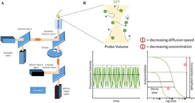
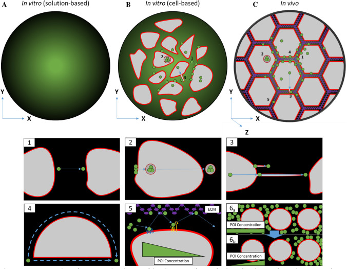
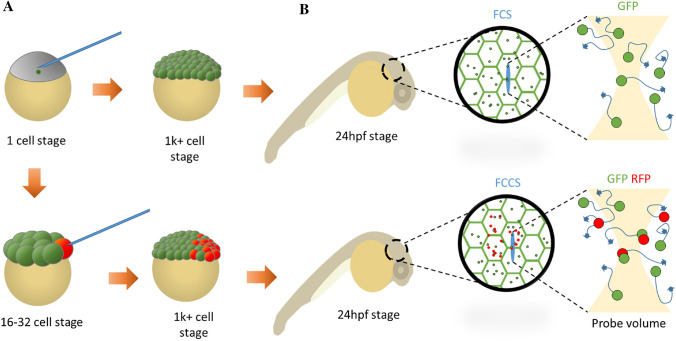
Cell behaviour and function is determined through the interactions of a multitude of molecules working in concert. To observe these molecular dynamics, biophysical studies have been developed that track single interactions. Fluorescence correlation spectroscopy (FCS) is an optical biophysical technique that non-invasively resolves single molecules through recording the signal intensity at the femtolitre scale. However, recording the behaviour of these biomolecules using in vitro-based assays often fails to recapitulate the full range of variables in vivo that directly confer dynamics. Therefore, there has been an increasing interest in observing the state of these biomolecules within living organisms such as the zebrafish Danio rerio. In this review, we explore the advancements of FCS within the zebrafish and compare and contrast these findings to those found in vitro.

Keywords: Fluorescent correlation spectroscopy (FCS); Microscopy; Protein–protein interactions; Zebrafish.


Figures

Fig. 1
Fig. 1
Overview of FCS set-up and zebrafish measurements. a A schematic of a typical FCS set-up. Single or dual excitation lasers can be configured to excite one or two types of fluorophores for FCS and fluorescence cross-correlation spectroscopy (FCCS), respectively. Dichroic mirrors are in place to split and/or reflect beam paths of certain wavelengths. The excitation beam is sent through the objective to excite fluorophores in a zebrafish embryo. Emission is detected back along the same optical path and through the excitation dichroic mirror. Long- and short-wavelength emissions are split by an emission dichroic mirror to allow avalanche photodiodes (APDs) to detect specific fluorophores. b Cartoon of probe volume that FCS laser beam passes through. The probe volume excites diffusing green fluorescent protein (GFP) and receives an emission spectrum. Emission beam signal intensity is recorded over time and transformed via a suitable fitting model into interpretable data that details the sample’s diffusion coefficient and concentration. (1) Reduced diffusion speed, (2) reduced concentration
Fig. 2
Fig. 2
Comparison between in vitro and in vivo sample analysis. a Solution-based assays rely on simple diffusion in an essentially homogenous solution. b Cell-based in vitro assays allow analysis of dynamics of intracellular processes such as (1) extracellular diffusion, (2) transcytosis and (3) the formation of signalling filopodia such as cytonemes. c In vivo (including three-dimensional collagen/matrigel cell-based) assays involve all aspects observed in cell-based in vitro assays and further parameters of molecular diffusion such as (4) tortuosity (hindrance of diffusion path by impermeable objects), (5) transient binding of molecules to the extracellular matrix (ECM) and the cell membrane, and (6) restrictive clearance for gradient formation and the additional Z-axis that introduces further dimensions for diffusion
Fig. 3
Fig. 3
mRNA injection time determines distribution of fluorophore. a Microinjection at very early embryo stage (1–4 cell) generates homogenous expression of fluorophore across entire embryo, while later stage injections (16–32 cell) generates confined/mosaic patterning of fluorophore. b Depending on time of injection, patterning on embryo can be imaged with FCS or with FCCS using two or more fluorophores

References

    1. Amemiya CT, et al. Zebrafish YAC, BAC, and PAC genomic libraries. Methods Cell Biol. 1999;60:235–258. - PubMed
    1. Anne SL, et al. WNT3 inhibits cerebellar granule neuron progenitor proliferation and medulloblastoma formation via MAPK activation. PLoS ONE. 2013;8(11):e81769. - PMC - PubMed
    1. Bai Y, et al. Ror2 receptor mediates Wnt11 ligand signaling and affects convergence and extension movements in zebrafish. J Biol Chem. 2014;289(30):20664–20676. - PMC - PubMed
    1. Beam M, Silva MC, Morimoto RI. Dynamic imaging by fluorescence correlation spectroscopy identifies diverse populations of polyglutamine oligomers formed in vivo. J Biol Chem. 2012;287(31):26136–26145. - PMC - PubMed
    1. Böttcher RT, Niehrs C. Fibroblast growth factor signaling during early vertebrate development. Endocr Rev. 2005;26(1):63–77. - PubMed

LinkOut - more resources