. 2021 Mar;23(1):184-198.
doi: 10.1007/s12017-020-08620-4. Epub 2020 Oct 16.

Effect of Ergothioneine on 7-Ketocholesterol-Induced Endothelial Injury

Affiliations

Effect of Ergothioneine on 7-Ketocholesterol-Induced Endothelial Injury

Sally Shuxian Koh et al. Neuromolecular Med. 2021 Mar.

Abstract

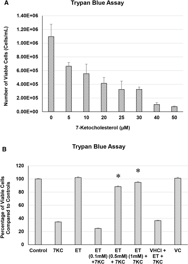
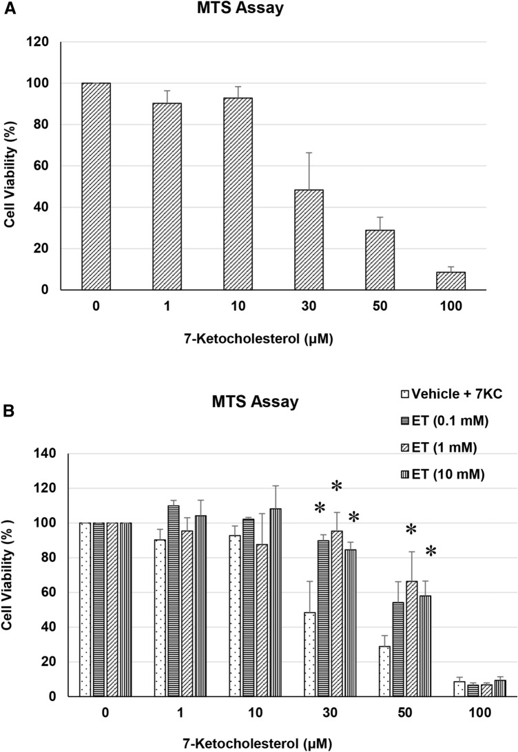
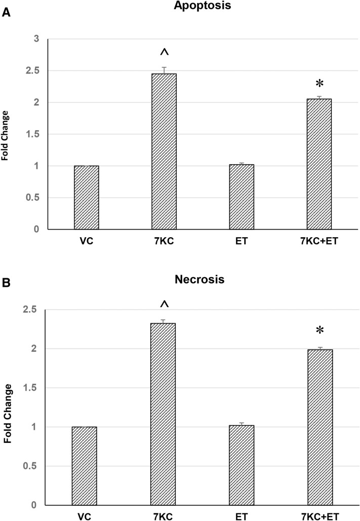
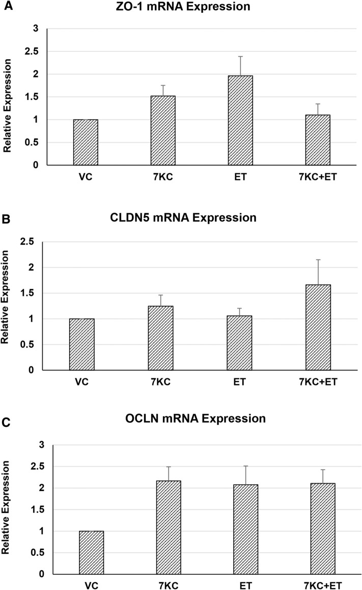
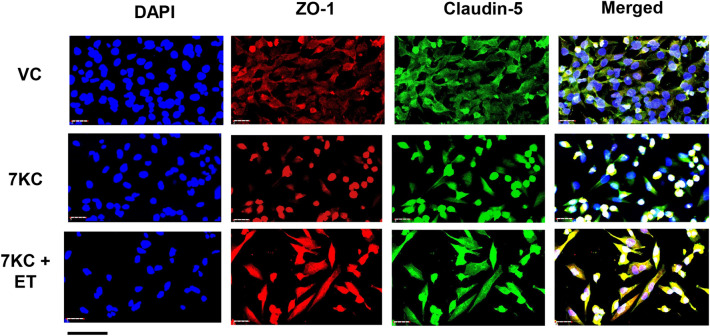
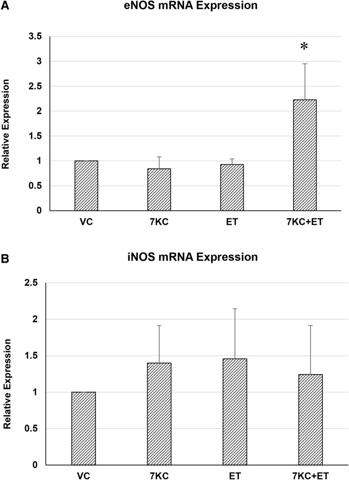
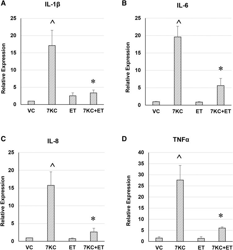
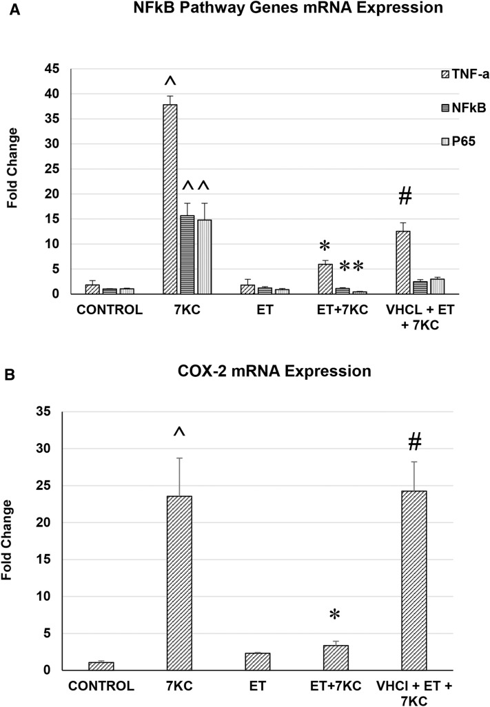
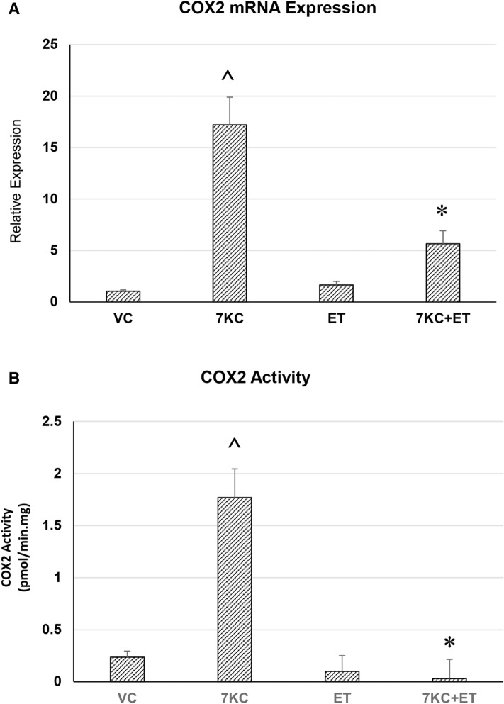
Ergothioneine (ET) is a naturally occurring antioxidant that is synthesized by non-yeast fungi and certain bacteria. ET is not synthesized by animals, including humans, but is avidly taken up from the diet, especially from mushrooms. In the current study, we elucidated the effect of ET on the hCMEC/D3 human brain endothelial cell line. Endothelial cells are exposed to high levels of the cholesterol oxidation product, 7-ketocholesterol (7KC), in patients with cardiovascular disease and diabetes, and this process is thought to mediate pathological inflammation. 7KC induces a dose-dependent loss of cell viability and an increase in apoptosis and necrosis in the endothelial cells. A relocalization of the tight junction proteins, zonula occludens-1 (ZO-1) and claudin-5, towards the nucleus of the cells was also observed. These effects were significantly attenuated by ET. In addition, 7KC induces marked increases in the mRNA expression of pro-inflammatory cytokines, IL-1β IL-6, IL-8, TNF-α and cyclooxygenase-2 (COX2), as well as COX2 enzymatic activity, and these were significantly reduced by ET. Moreover, the cytoprotective and anti-inflammatory effects of ET were significantly reduced by co-incubation with an inhibitor of the ET transporter, OCTN1 (VHCL). This shows that ET needs to enter the endothelial cells to have a protective effect and is unlikely to act via extracellular neutralizing of 7KC. The protective effect on inflammation in brain endothelial cells suggests that ET might be useful as a nutraceutical for the prevention or management of neurovascular diseases, such as stroke and vascular dementia. Moreover, the ability of ET to cross the blood-brain barrier could point to its usefulness in combatting 7KC that is produced in the CNS during neuroinflammation, e.g. after excitotoxicity, in chronic neurodegenerative diseases, and possibly COVID-19-related neurologic complications.

Keywords: 7-ketocholesterol; Anti-inflammatory; Antioxidant; Blood–brain barrier; COVID-19; COX2; Claudin-5; Coronavirus; Cyclooxygenase; Cytokines; Eicosanoids; Ergothioneine; Free radicals; Inflammation; Mushrooms; NF-kB; Oxidative stress; Oxysterols; TNF-α; Tight junction proteins; ZO-1.


Conflict of interest statement

The authors have no conflicts of interest to declare.

Figures

Fig. 1
Fig. 1
a Effect of 7KC on endothelial cells in culture, determined using the trypan blue dye exclusion assay. a dose-dependent cytotoxicity of 7KC. hCMEC/D3 cells were treated with at 0–50 μM 7KC for 24 h. Cell viability is expressed as percentage viability from Vehicle Control. The oxysterol induced significant loss in viable cells at 30-100 μM. Data are represented as mean ± S.E.M. (n = 4). b protective effect of ET on 7KC cytotoxicity. hCMEC/D3 cells were pre-treated with different concentrations of ET for 1 h, followed by co-treatment with 7KC for 24 h. Co-treatment of cells with ET results in modulation of 7KC-induced loss of viable cells. Data are represented as mean ± S.E.M. (n = 4). * Significant difference compared to 7KC (p < 0.01)
Fig. 2
Fig. 2
a Effect of 7KC on endothelial cells in culture, determined using the MTS assay. a dose-dependent cytotoxicity of 7KC. hCMEC/D3 cells were treated with at 0–100 μM 7KC for 24 h. Cell viability is expressed as percentage viability from Vehicle Control. The oxysterol induced significant loss in viable cells at 30–100 μM. Data are represented as mean ± S.E.M. (n = 3). b protective effect of ET on 7KC cytotoxicity. hCMEC/D3 cells were pre-treated with different concentrations of ET for 1 h, followed by co-treatment with 7KC for 24 h. Co-treatment of cells with ET results in modulation of 7KC-induced loss of viable cells. Data is represented as mean ± S.E.M. (n = 3). * Significant difference compared to 7KC (p < 0.01)
Fig. 3
Fig. 3
a Mode of cell death of hCMEC/D3 human brain endothelial cells after 30 μM 7KC and ET treatment, determined by flow cytometry. a Plot showing effect of 7KC. The oxysterol induced significant increase in percentage of cells that are labelled for phosphatidylserine, indicating apoptosis. At the same time, the oxysterol increased another population of cells that were labelled with 7-AAD, indicating necrosis. Yet a third population of cells showed neither the apoptotic nor necrotic marker. b ET by itself had no significant effect on apoptosis or necrosis; however, it significantly modulated the increase in apoptosis and necrosis after addition of 7KC. Data are represented as mean ± S.E.M. (n = 3). ^ Significant difference compared to controls. *Significant difference compared to 7KC (both p < 0.01)
Fig. 4
Fig. 4
Quantitative RT-PCR of Zonula occludens-1 (ZO-1), claudin-5 and occludin (OCLN) mRNA expression after 7KC and or ET treatment. hCMEC/D3 cells were pre-treated with 1 mM ET for 1 h, followed by co-treatment with 30 μM 7KC for 24 h. No significant changes in mRNA expression of endothelial tight junction proteins, ZO-1, claudin-5 or occludin were detected. Data are represented as mean ± S.E.M (n = 5)
Fig. 5
Fig. 5
Effect of 7KC and ET on cellular localization of blood–brain barrier tight junction proteins, determined by immunocytochemistry. hCMEC/D3 cells were pre-treated with 1 mM ET for 1 h, followed by co-treatment with 50 μM 7KC for 24 h. 7KC induced a movement of ZO-1 and claudin-5 towards the nucleus, but this change of distribution was abolished by ET. Scale bar = 40 μm
Fig. 6
Fig. 6
Quantitative RT-PCR of eNOS a or iNOS b mRNA expression after 7KC and /or ET treatment. hCMEC/D3 cells were pre-treated with 1 mM ET for 1 h, followed by co-treatment with 30 μM 7KC for 24 h. No significant changes in mRNA expression of these two NOS isoforms were detected. Data are represented as mean ± S.E.M (n = 4). * Significant difference compared to 7KC (p < 0.01)
Fig. 7
Fig. 7
Effect of ET on 7KC-induced increase in mRNA expression of pro-inflammatory genes, determined by real-time RT-PCR. hCMEC/D3 cells were pre-treated with 1 mM ET for 1 h, followed by co-treatment with 30 μM 7KC for 24 h. Co-treatment of cells with ET results in modulation of 7KC-induced increase in IL-1β (a), IL-6 (b), IL-8 (c) and TNFα (d) mRNA expression in endothelial cells. Data are represented as mean ± S.E.M. ^ Significant difference compared to controls. * Significant difference compared to 7KC (both p < 0.01) (n = 6)
Fig. 8
Fig. 8
Effect of ET on 7KC-induced increase in mRNA expression of pro-inflammatory genes, determined by real-time RT-PCR. hCMEC/D3 cells were pre-treated with 1 mM ET for 1 h, followed by co-treatment with 50 μM 7KC for 24 h. Co-treatment of cells with ET results in modulation of 7KC-induced increase in NFkB and component genes (a), as well as COX-2 (b) mRNA expression in endothelial cells. This effect was abolished by concurrent treatment with an inhibitor of ET transporter, VHCl. Data are represented as mean ± S.E.M. (n = 4). ^ Significant difference compared to controls. * Significant difference compared to 7KC. # Significant difference compared to ET + 7KC (all p < 0.01)
Fig. 9
Fig. 9
Effect of ET on 7KC-induced increase in COX-2 mRNA expression (A) and COX-2 enzymatic activity (B) hCMEC/D3 cells were pre-treated with 1 mM ET for 1 h, followed by co-treatment with 50 μM 7KC for 24 h. Co-treatment of cells with ET results in modulation of 7KC-induced increase in COX-2 expression and activity. Data are represented as mean ± S.E.M. (n = 4). ^ Significant difference compared to controls. * Significant difference compared to 7KC (all p < 0.01)

References

    1. Akanmu D, Cecchini R, Aruoma OI, Halliwell B. The antioxidant action of ergothioneine. Archives of Biochemistry and Biophysics. 1991;288(1):10–16. doi: 10.1016/0003-9861(91)90158-f. - DOI - PubMed
    1. Anderson A, Campo A, Fulton E, Corwin A, Jerome WG, 3rd, O'Connor MS. 7-Ketocholesterol in disease and aging. Redox Biol. 2020;29:101380. doi: 10.1016/j.redox.2019.101380. - DOI - PMC - PubMed
    1. Anticoli S, Arciello M, Mancinetti A, De Martinis M, Ginaldi L, Iuliano L, et al. 7-ketocholesterol and 5,6-secosterol modulate differently the stress-activated mitogen-activated protein kinases (MAPKs) in liver cells. Journal of Cellular Physiology. 2010;222(3):586–595. doi: 10.1002/jcp.21972. - DOI - PubMed
    1. Brown AJ, Watts GF, Burnett JR, Dean RT, Jessup W. Sterol 27-hydroxylase acts on 7-ketocholesterol in human atherosclerotic lesions and macrophages in culture. Journal of Biological Chemistry. 2000;275(36):27627–27633. doi: 10.1074/jbc.M004060200. - DOI - PubMed
    1. Butterfield DA, Halliwell B. Oxidative stress, dysfunctional glucose metabolism and Alzheimer disease. Nature Reviews Neuroscience. 2019;20(3):148–160. doi: 10.1038/s41583-019-0132-6. - DOI - PMC - PubMed

Publication types

MeSH terms

Substances