. 2021 Feb 15;42(3):567-586.
doi: 10.1002/hbm.25242. Epub 2020 Oct 17.

Construction and validation of a database of head models for functional imaging of the neonatal brain

Affiliations

Construction and validation of a database of head models for functional imaging of the neonatal brain

Liam H Collins-Jones et al. Hum Brain Mapp. .

Abstract

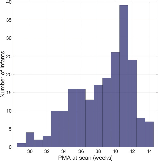
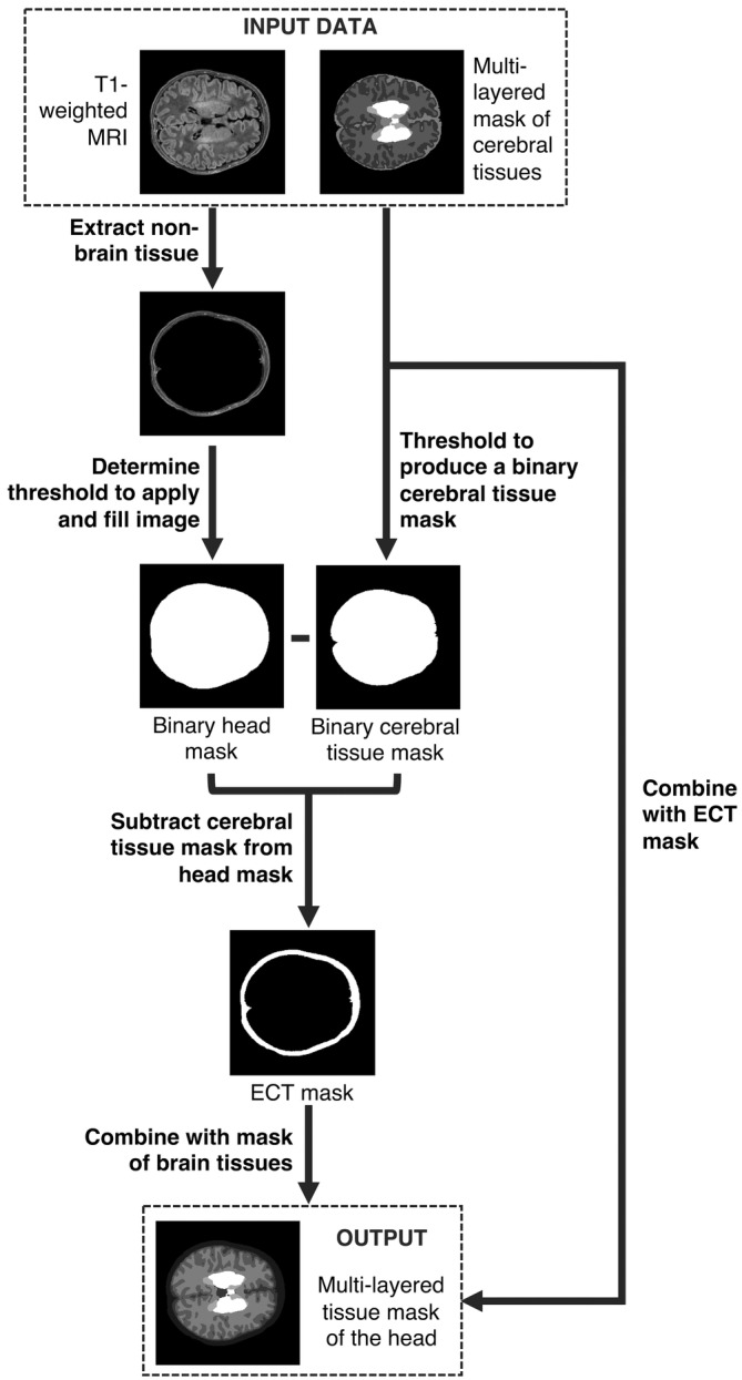
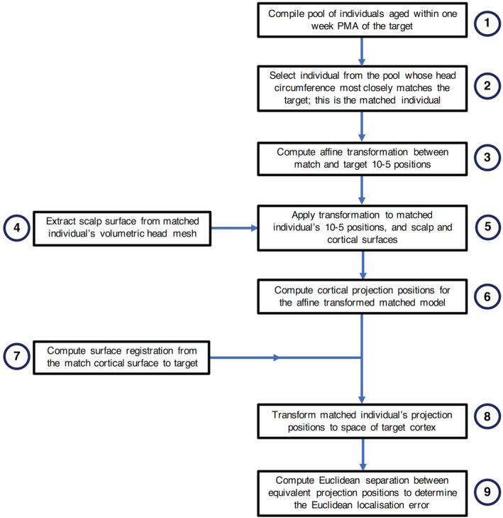
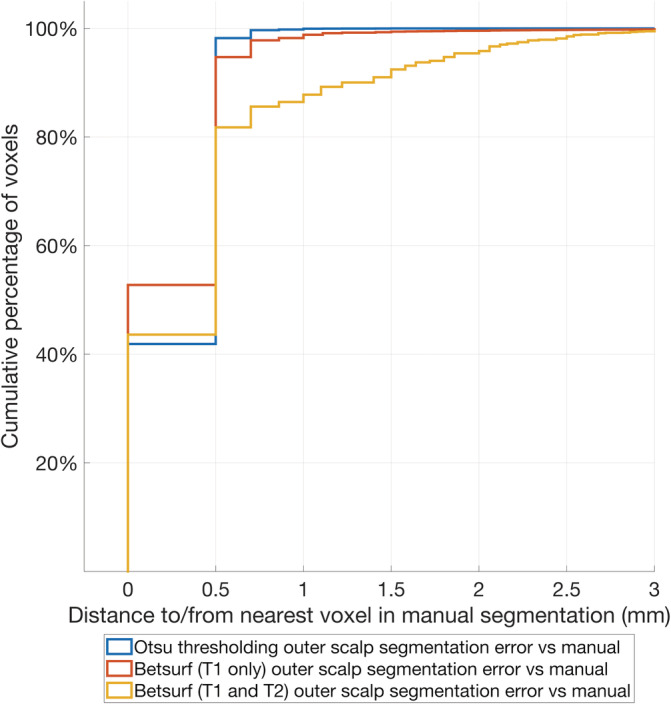
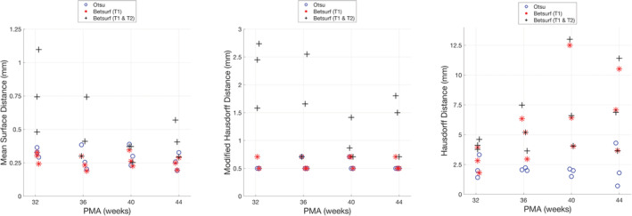
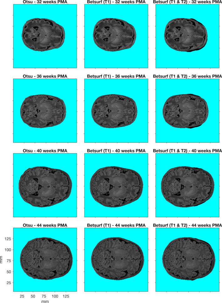
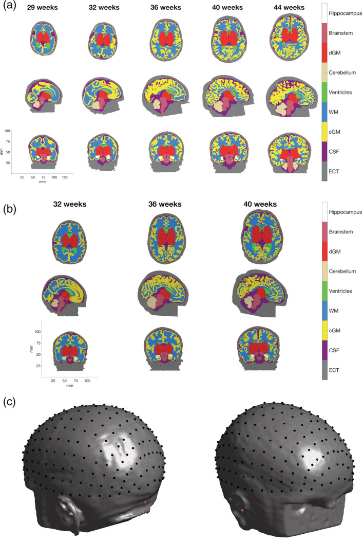
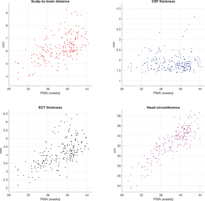
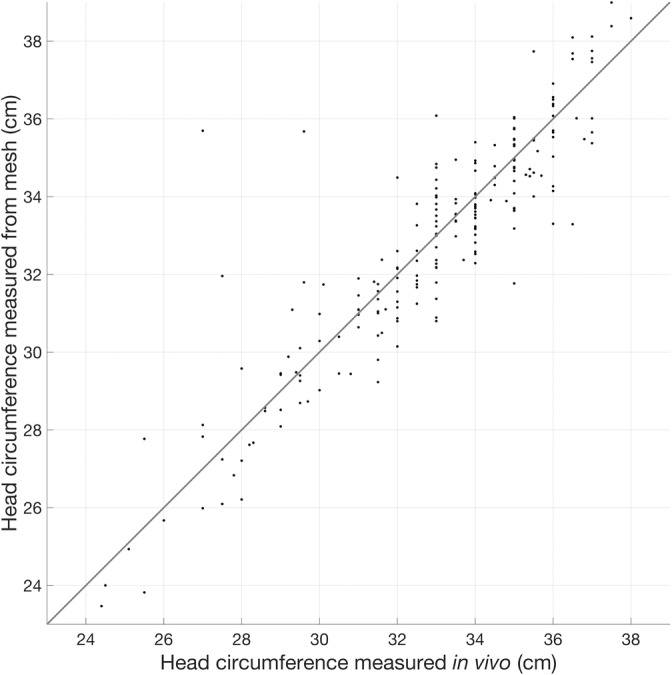
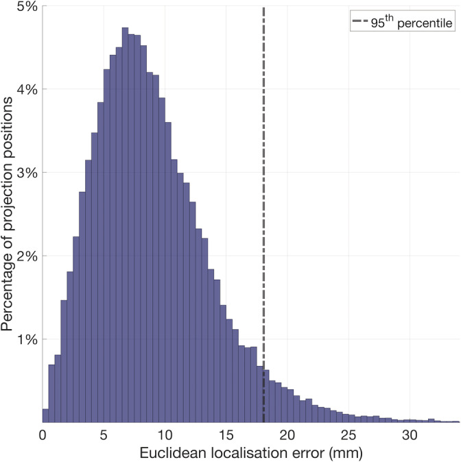
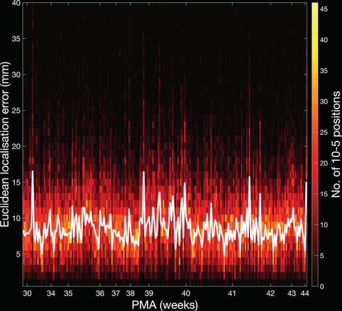
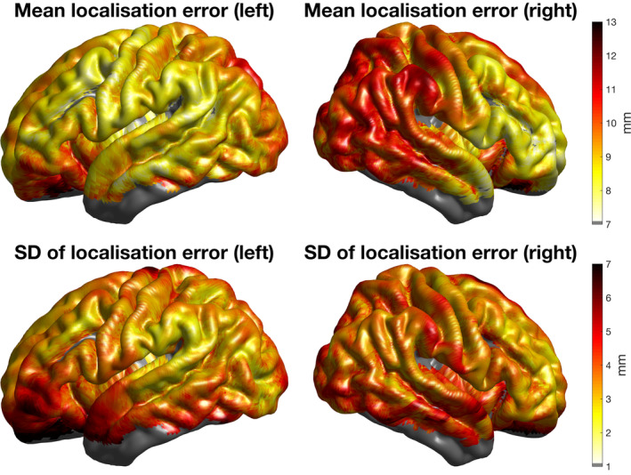
The neonatal brain undergoes dramatic structural and functional changes over the last trimester of gestation. The accuracy of source localisation of brain activity recorded from the scalp therefore relies on accurate age-specific head models. Although an age-appropriate population-level atlas could be used, detail is lost in the construction of such atlases, in particular with regard to the smoothing of the cortical surface, and so such a model is not representative of anatomy at an individual level. In this work, we describe the construction of a database of individual structural priors of the neonatal head using 215 individual-level datasets at ages 29-44 weeks postmenstrual age from the Developing Human Connectome Project. We have validated a method to segment the extra-cerebral tissue against manual segmentation. We have also conducted a leave-one-out analysis to quantify the expected spatial error incurred with regard to localising functional activation when using a best-matching individual from the database in place of a subject-specific model; the median error was calculated to be 8.3 mm (median absolute deviation 3.8 mm). The database can be applied for any functional neuroimaging modality which requires structural data whereby the physical parameters associated with that modality vary with tissue type and is freely available at www.ucl.ac.uk/dot-hub.

Keywords: atlas; database; neonatal; structural prior.


Conflict of interest statement

The authors declare no conflicts of interest.

Figures

FIGURE 1
FIGURE 1
Age distribution of infants whose structural data were used to produce the database of neonatal structural priors
FIGURE 2
FIGURE 2
Flowchart describing the construction of structural priors for each neonatal individual using segmentations of cerebral tissues and an extra‐cerebral tissue (ECT) segmentation extracted from a T1‐weighted MR image
FIGURE 3
FIGURE 3
Flowchart of the process to compute the Euclidean localisation error for each individual acting as the target in a leave‐one‐out paradigm
FIGURE 4
FIGURE 4
Cumulative distribution plot of the distances to/from the centre of each manually segmented boundary voxel from/to the centre of the nearest automated segmentation boundary voxel for each of the three automated methods. These distances are used as a measure of the error of the outer scalp boundary relative to manual segmentation
FIGURE 5
FIGURE 5
The metrics used to evaluate the automated segmentation methods are shown for each infant in the subset evaluated with manual segmentation
FIGURE 6
FIGURE 6
T1‐weighted images from example individuals at 32, 36, 40, and 44 weeks postmenstrual age (PMA). For each individual, the outer scalp boundaries determined using three different automated segmentation methods are shown (demarcated by the turquoise background)
FIGURE 7
FIGURE 7
Example of multi‐layered (a) tissue masks and (b) meshes from neonatal infants aged 29–44 weeks postmenstrual age (PMA) (see colourbar). Tissues represented are extra‐cerebral tissue (ECT), cerebrospinal fluid (CSF), cortical grey matter (cGM), white matter (WM), ventricles, cerebellum, deep grey matter (dGM), brainstem, and hippocampus. The 10–5 positions on the scalp surface from an example infant aged 41 weeks PMA are shown in (c) in black, while the cranial landmarks are shown in magenta
FIGURE 8
FIGURE 8
Median tissue thickness values underlying the 10–5 positions and head circumference as a function of age for all individuals in the database
FIGURE 9
FIGURE 9
Head circumference measurements taken in vivo (x‐axis) plotted against head circumference measurements taken from volumetric meshes (y‐axis). The line of one‐to‐one proportion is shown
FIGURE 10
FIGURE 10
Histogram displaying the distribution of the Euclidean localisation error of each cortical projection position, combining data from each individual
FIGURE 11
FIGURE 11
Histogram displaying the distribution of the Euclidean localisation error of the cortical projection positions per individual (see colourbar) and the median values for each individual (plotted as a white line)
FIGURE 12
FIGURE 12
For each individual aged 41 weeks postmenstrual age (PMA), the Euclidean localisation error was interpolated across the cortical surface using the value at each projection position. The interpolated localisation error maps for each of these individuals were registered to the surface of a 41‐week PMA cortical surface atlas and averaged. Here, the node‐wise values of the mean and SD are displayed

References

    1. Aasted, C. M. , Yücel, M. A. , Cooper, R. J. , Dubb, J. , Tsuzuki, D. , Becerra, L. , … Boas, D. A. (2015). Anatomical guidance for functional near‐infrared spectroscopy: AtlasViewer tutorial. Neurophotonics, 2(2), 020801 10.1117/1.nph.2.2.020801 - DOI - PMC - PubMed
    1. Alexander, B. , Murray, A. L. , Loh, W. Y. , Matthews, L. G. , Adamson, C. , Beare, R. , … Thompson, D. K. (2017). A new neonatal cortical and subcortical brain atlas: The Melbourne Children's Regional Infant Brain (M‐CRIB) atlas. NeuroImage, 147, 841–851. 10.1016/j.neuroimage.2016.09.068 - DOI - PubMed
    1. Allievi, A. G. , Arichi, T. , Tusor, N. , Kimpton, J. , Arulkumaran, S. , Counsell, S. J. , … Burdet, E. (2016). Maturation of sensori‐motor functional responses in the preterm brain. Cerebral Cortex, 26(1), 402–413. 10.1093/cercor/bhv203 - DOI - PMC - PubMed
    1. Arichi, T. , Moraux, A. , Melendez, A. , Doria, V. , Groppo, M. , Merchant, N. , … Edwards, A. D. (2010). Somatosensory cortical activation identified by functional MRI in preterm and term infants. NeuroImage, 49(3), 2063–2071. 10.1016/j.neuroimage.2009.10.038 - DOI - PubMed
    1. Arichi, T. , Fagiolo, G. , Varela, M. , Melendez‐Calderon, A. , Allievi, A. , Merchant, N. , … Edwards, A. D. (2012). Development of BOLD signal hemodynamic responses in the human brain. NeuroImage, 63(2), 663–673. 10.1016/j.neuroimage.2012.06.054 - DOI - PMC - PubMed

Publication types