Review
. 2021 Jan 1:519:111052.
doi: 10.1016/j.mce.2020.111052. Epub 2020 Oct 14.

Effects of GH/IGF axis on bone and cartilage

Affiliations
Review

Effects of GH/IGF axis on bone and cartilage

Manisha Dixit et al. Mol Cell Endocrinol. .

Abstract

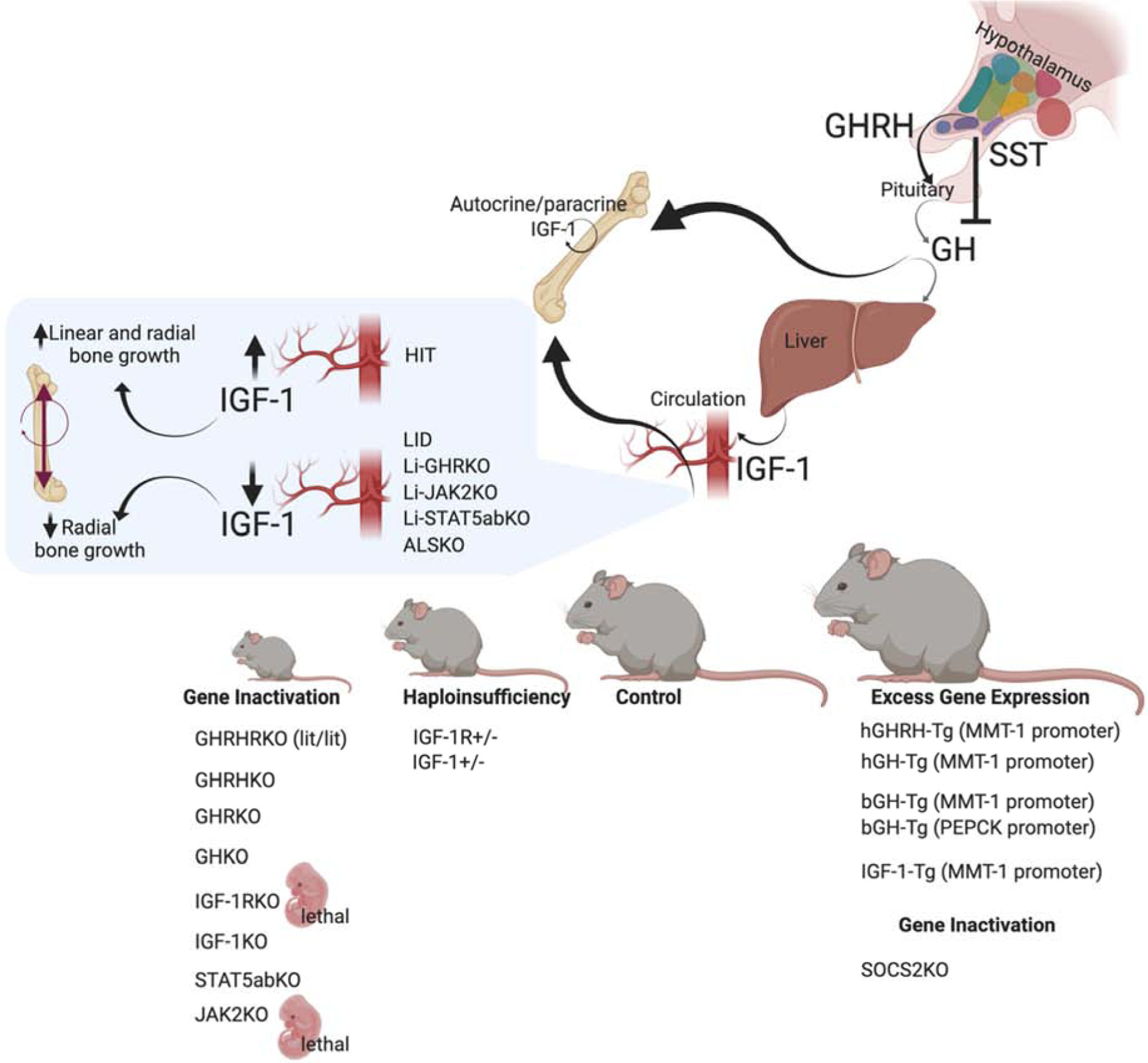
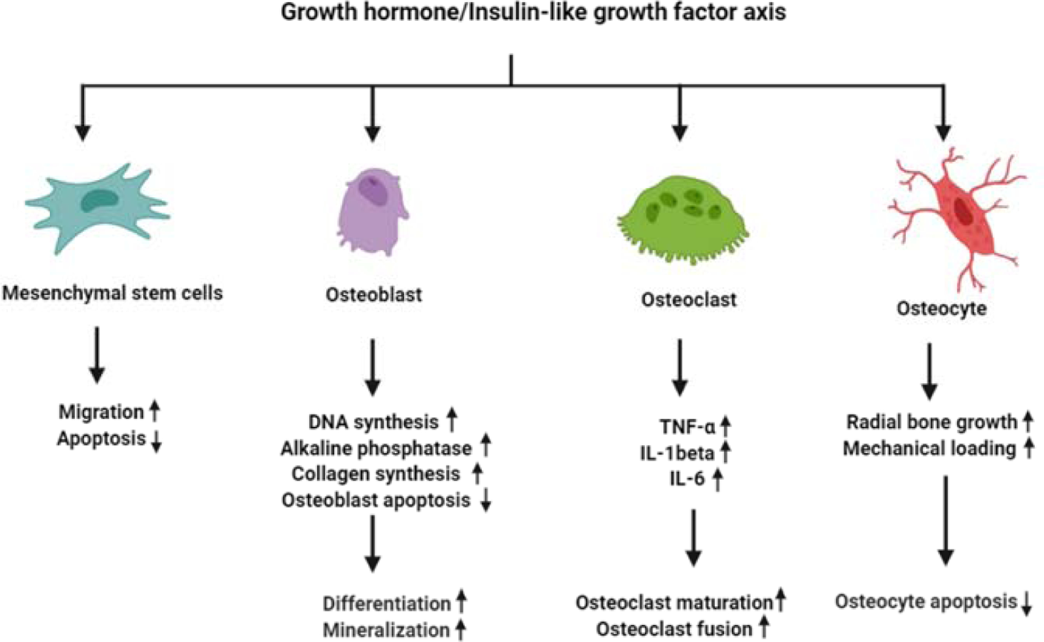
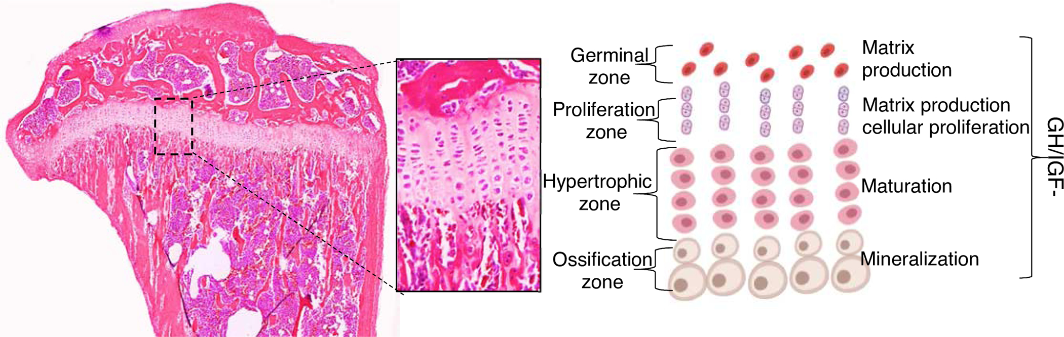
Growth hormone (GH) and its mediator, the insulin-like growth factor-1 (IGF-1) regulate somatic growth, metabolism and many aspects of aging. As such, actions of GH/IGF have been studied in many tissues and organs over decades. GH and IGF-1 are part of the hypothalamic/pituitary somatotrophic axis that consists of many other regulatory hormones, receptors, binding proteins, and proteases. In humans, GH/IGF actions peak during pubertal growth and regulate skeletal acquisition through stimulation of extracellular matrix production and increases in bone mineral density. During aging the activity of these hormones declines, a state called somatopaguss, which associates with deleterious effects on the musculoskeletal system. In this review, we will focus on GH/IGF-1 action in bone and cartilage. We will cover many studies that have utilized congenital ablation or overexpression of members of this axis, as well as cell-specific gene-targeting approaches used to unravel the nature of the GH/IGF-1 actions in the skeleton in vivo.

Keywords: Bone; Cartilage; Growth hormone; Insulin-like growth factor-1.


Figures

Figure 1:
Figure 1:. Mouse models of the GH/IGF axis.
Several naturally occurring mutations in the GH/IGF-1 axis have been described over the years. Among them is the lit/lit mouse line, with a mutation in the GHRH-receptor which causes pituitary hypoplasia, reduced GH secretion, and dwarfism [220]. Similarly, targeted ablation of the GHRH gene causes GH deficiency, pituitary hypoplasia, and reductions in somatic growth [221]. GHRKO and the recently described GHKO mice [222] also show decreased linear bone growth and reduced whole-body BMD. Gene ablation of the IGF-1R was lethal [57], and caused severely retarded growth, with delayed ossification in all bones. Likewise, IGF-1KO mice showed severe dwarfism, associated with reduced BMD and decreased linear and radial bone growth [223]. In mice, inactivation of the downstream mediator of GH signaling, STAT5ab, also affected linear growth [224]. However, the details of skeletal morphology were not reported. Finally, in mice, knocking out JAK2, the GH mediator, was lethal [225], likely due to its central roles in cytokine signaling. Haploinsufficiency of IGF-1 or the IGR-1R associate with intermediate growth retardation and reduced linear and radial bone growth. Mouse models of excess GHRH [35, 226], hGH [–42], and bGH [, –49] show increased somatic growth with robust bones. Mice with excess IGF-1 resulted in selective organomegaly and as reported at the time showed minimal effects on skeletal growth [52, 227]. Lastly, SOCS2KO mice, which exhibit a “physiological” excess of GH action (as oppose to transgene driven GH expression), show increased linear and radial bone growth with increased trabecular bone volume [228].
Figure 2:
Figure 2:. The effects of GH and IGF-1 on cells of the osseous system.
GH and IGF-1 stimulate the proliferation of MSCs and osteoprogenitors, and promote their differentiation into OB and chondrocytes. Activation of the IGF-1 and GH receptors in OB leads to increased OB proliferation, as evidenced by increased DNA synthesis. It also leads to OB cell differentiation, which is associated with increased collagen synthesis and elevated alkaline-phosphatase activity. Additionally, a mouse model of targeted inactivation of the IGF-1R in OB revealed the significant roles played by IGF-1 in matrix mineralization. Bone remodeling requires coupling of OB and OCL activity. Several mouse models have shown that IGF-1 also affects OCL maturation, fusion, and activity. The communication between OBs and OCLs is believed to be mediated by IGF-dependent expression of the membrane-bound ligand/receptor system ephrinB2/Eph4. Finally, GH IGF-1 receptors on osteocytes are important in regulation of radial bone growth and play significant roles in bone response to mechanical loading (not discussed in the current review).
Figure 3:
Figure 3:. The growth-plate.
The effects of the GH/IGF axis on chondrocytes of the growth plate were extensively studied (and described in the current review). GH elicits direct and IGF-dependent effects on chondrocytes in all zones of the growth plate. However, IGF-1 actions were mostly detected at the proliferative zone, while those of GH were localized to the germinal zone.
Figure 4:
Figure 4:. The effects of GH/IGF-1 axis on OA initiation and progression.
OA is characterized by a progressive degeneration of the articular cartilage, leading to changes in the entire synovial joint structure. These include narrowing of the joint space, sinovitis, deterioration of subchondral bone volume and structure, and formation of osteophytes. The roles of the GH/IGF-1 axis in OA are not fully understood. However, studies have shown a bell-shaped relationship between OA and GH/IGF signals, where too-low or too-high levels of these two hormones are associated with the joint disease.

References

    1. Tuggle CK and Trenkle A, Control of growth hormone synthesis. Domest Anim Endocrinol, 1996. 13(1): p. 1–33. - PubMed
    1. Gahete MD, et al. , Understanding the multifactorial control of growth hormone release by somatotropes: lessons from comparative endocrinology. Ann N Y Acad Sci, 2009. 1163: p. 137–53. - PubMed
    1. Pincus SM, et al. , Females secrete growth hormone with more process irregularity than males in both humans and rats. Am J Physiol, 1996. 270(1 Pt 1): p. E107–15. - PubMed
    1. Spiess J, Rivier J, and Vale W, Characterization of rat hypothalamic growth hormone-releasing factor. Nature, 1983. 303(5917): p. 532–5. - PubMed
    1. Brazeau P, et al. , Hypothalamic polypeptide that inhibits the secretion of immunoreactive pituitary growth hormone. Science, 1973. 179(4068): p. 77–9. - PubMed

Publication types

Substances