. 2020 Oct 17;18(1):144.
doi: 10.1186/s12951-020-00707-1.

Interaction kinetics of peptide lipids-mediated gene delivery

Affiliations

Interaction kinetics of peptide lipids-mediated gene delivery

Yinan Zhao et al. J Nanobiotechnology. .

Abstract

Background: During the course of gene transfection, the interaction kinetics between liposomes and DNA is speculated to play very important role for blood stability, cellular uptake, DNA release and finally transfection efficiency.

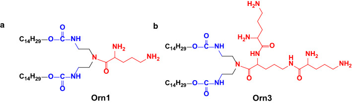
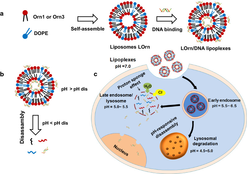
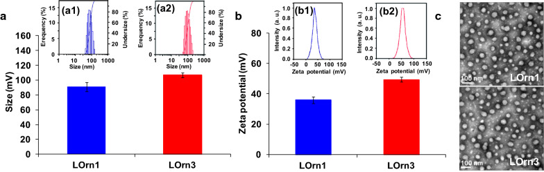
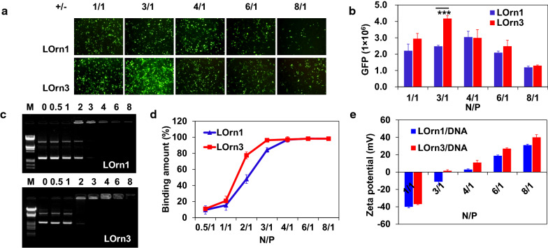
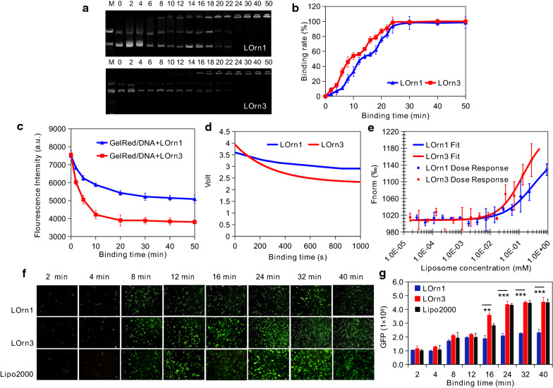
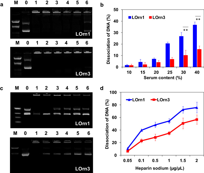
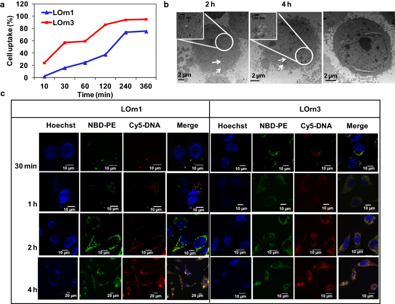
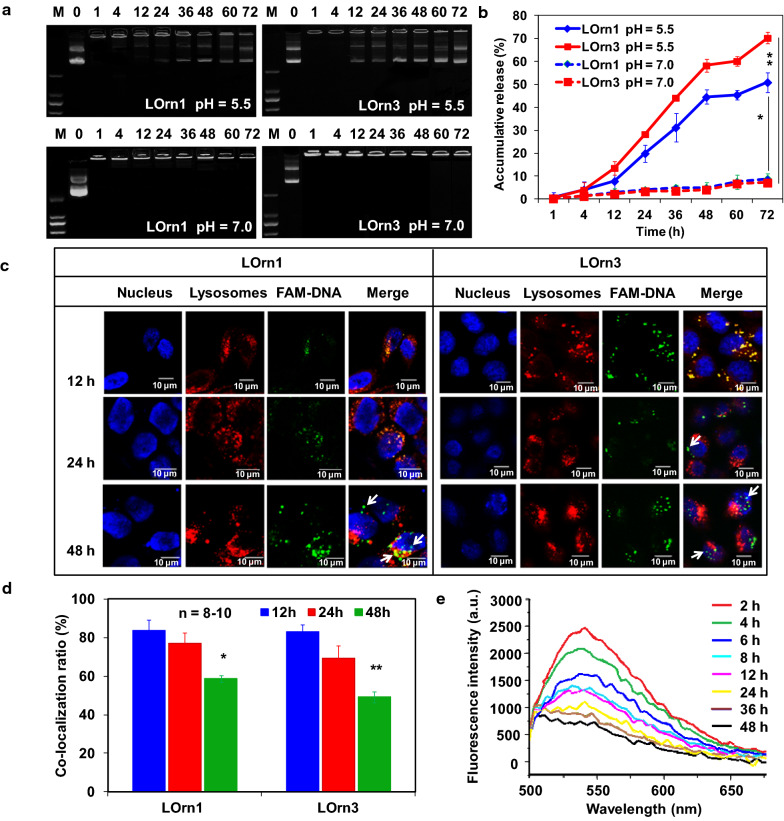
Results: As cationic peptide liposomes exhibited great gene transfer activities both in vitro and in vivo, two peptide lipids, containing a tri-ornithine head (LOrn3) and a mono-ornithine head (LOrn1), were chosen to further clarify the process of liposome-mediated gene delivery in this study. The results show that the electrostatically-driven binding between DNA and liposomes reached nearly 100% at equilibrium, and high affinity of LOrn3 to DNA led to fast binding rate between them. The binding process between LOrn3 and DNA conformed to the kinetics equation: y = 1.663631 × exp (- 0.003427x) + 6.278163. Compared to liposome LOrn1, the liposome LOrn3/DNA lipoplex exhibited a faster and more uniform uptake in HeLa cells, as LOrn3 with a tri-ornithine peptide headgroup had a stronger interaction with the negatively charged cell membrane than LOrn1. The efficient endosomal escape of DNA from LOrn3 lipoplex was facilitated by the acidity in late endosomes, resulting in broken carbamate bonds, as well as the "proton sponge effect" of the lipid.

Conclusions: The interaction kinetics is a key factor for DNA transfection efficiency. This work provided insights into peptide lipid-mediated DNA delivery that could guide the development of the next generation of delivery systems for gene therapeutics.

Keywords: Gene delivery; Gene release; Interaction kinetics; Peptide lipids; Uptake.


Conflict of interest statement

The authors declare that they have no competing interests.

Figures

Fig. 1
Fig. 1
Structures of the peptide lipids (a) Orn1 and (b) Orn3
Fig. 2
Fig. 2
Assembling process of the liposome/DNA lipoplex and the intracellular transport of internalized DNA. a Self-assembly of a liposome and the interaction of a liposome with DNA. b Dissociation of a lipoplex. c Lipoplexes respond to the slightly acidic environment of early endosomes and release the cargoes
Fig. 3
Fig. 3
Characterization of liposomes. a Particle size and (b) Zeta potential of liposomes measured by using a ZetaSizer. Particle size distribution of LOrn1and LOrn3 is shown in (a1, a2), the PDI of LOrn1 and LOrn3 was 0.102 ± 0.014 and 0.097 ± 0.006, respectively. Zeta potential distribution of LOrn1 and LOrn3 is shown in (b1, b2). c Morphology of the particles measured by TEM, the diameters of Lorn1 and LOrn3 were 42.85 ± 16.3 nm and 44.16 ± 12.07 nm, respectively (voltage: 100 kV)
Fig. 4
Fig. 4
a Transfection efficiency of lipoplexes against HeLa cells. GFP expression of pGFP-N1 was mediated by liposomes LOrn1 and LOrn3 at the charge ratios ( ±) of 1:1, 3:1, 4:1, 6:1 and 8:1. The measurement was carried out in cells by using an inverted fluorescence microscope (10 × 10). b Transfection efficiency in Figure (a) was quantified by flow cytometry analysis. c Agarose gel electrophoresis of liposomes/DNA lipoplexes at various charge ratios. Line M: marker [λ DNA/EcoRI + HindIII]; Line 0: naked DNA [0.2 μg]; Lines 0.5–8: charge ratios were 0.5:1, 1:1, 2:1, 3:1, 4:1, 6:1 and 8:1. d Quantitative analysis by automatic analysis system of gel imaging. e Zeta potential of liposomes/DNA lipoplexes at charge ratios of 1:1, 3:1, 4:1, 6:1 and 8:1. Significance levels: ***p < 0.001
Fig. 5
Fig. 5
Kinetic traces for binding of peptide liposome and DNA. a Agarose gel electrophoresis of the lipoplexes at charge ratios of 4:1 and 3:1 for lipids LOrn1 and LOrn3, respectively (Line M: marker [λ DNA/EcoRI + HindIII]; Line 0: naked DNA [0.2 μg]; Lines 2–50: binding times were 2, 4, 6, 8, 10, 12, 14, 16, 18, 20, 22, 24, 30, 40 and 50 min). b Quantitative analysis by automatic analysis system of gel imaging. c Fluorescence intensity of labeled DNA with GelRed and liposomes bound to DNA at charge ratio ( ±) 3:1, at several interaction times: 0, 2, 5, 10, 20, 30, 40, and 50 min. d Stopped-flow fluorescence intensity decay of LOrn1/DNA and LOrn3/DNA lipoplexes binding at the charge ratios of 4:1 and 3:1, respectively. The final DNA and GelRed concentrations were 2 µg/mL and 0.2 µL/mL, respectively. The fluorescence was measured by excitation at a wavelength of 260 nm. e Affinity of liposomes to DNA, Kd values of LOrn1 and LOrn3 were 0.296 and 0.136 µM, respectively. All values are expressed as mean ± SD (n = 5). f Effect of liposome-DNA binding time on the transfection efficiency of liposomes with 2, 4, 8, 12, 16, 24, 32 and 40 min. g Quantitative analysis in Figure (f) by flow cytometry. All values are expressed as mean ± SD (n = 5). The amount of pGFP-N1 was 1 µg/well for 24-well plates. Lipo2000 was used as control. Significance levels: **p < 0.01, ***p < 0.001
Fig. 6
Fig. 6
Effect of serum and heparin sodium on the stability of LOrn1/DNA lipoplexes with a charge ratio of 4:1 and LOrn3/DNA lipoplexes with an charge ratio of 3:1. a Agarose gel electrophoresis of the lipoplexes at different serum contents (Lane M: marker; Lane DNA: free DNA; Lanes 1–6: serum contents of 10%, 15%, 20%, 25%, 30%, and 40%, respectively). b Quantitative analysis of the dissociation rate of lipoplexes in the present of serum. c Agarose gel electrophoresis of the lipoplexes at different concentrations of heparin sodium (Lane M: marker; Lane DNA: free DNA; Lanes 1–6: heparin sodium concentrations of 0.05, 0.1, 0.5, 1.0, 1.5, and 2.0 µg/µL, respectively). d Quantitative analysis of the dissociation rate of lipoplexes in the present of heparin sodium. Each bar represents the mean ± SD (n = 5). Significance levels: **p < 0.01
Fig. 7
Fig. 7
a Cellular uptake of liposome/FAM-DNA lipoplex in HeLa cells was evaluated by FACS. b Intracellular trafficking of LOrn3/DNA lipoplex in HeLa cells at incubation time of 30 min, 2 h, and 4 h by TEM. c Intracellular tracking of lipoplexes with incubation time of 30 min, 1 h, 2 h, and 4 h in HeLa cells by LSCM. Hoechst was used to label cell nucleus (blue), NBD-PE to label liposomes (green), and Cy5 to label DNA (red)
Fig. 8
Fig. 8
a DNA dissociation profile of the LOrn1/DNA and LOrn3/DNA lipoplexes, evaluated by gel electrophoresis at a pH of 5.5; the lipoplexes were measured at a pH of 7.0 as controls. b Quantitative analysis of accumulative DNA release by gel electrophoresis. Significance levels: *p < 0.05, **p < 0.01. c Intracellular release of DNA in HeLa cells as determined by LSCM. White arrows showed the aggregation state of FAM-labeled DNA. d Co-localization ratio of FAM-DNA (green) and lysosomes (red). 8–10 Cells in each group were selected randomly. Significance levels: *p < 0.05 vs. 12 h, **p < 0.01 vs. 12 h. e Fluorescence spectra of Rhodamine 123 in HeLa cell lysates with culture times of 2, 4, 6, 8, 12, 24, 36, and 48 h after transfection with LOrn3/DNA lipoplex

References

    1. Cavazzana-Calvo M, Thrasher A, Mavilio F. The future of gene therapy. Nature. 2004;427:779–781. doi: 10.1038/427779a. - DOI - PubMed
    1. Kaiser J. Gene therapists celebrate a decade of progress. Science. 2011;334:29–30. doi: 10.1126/science.334.6052.29. - DOI - PubMed
    1. Niu J, Chu Y, Huang YF, Chong YS, Jiang ZH, Mao ZW, Peng LH, Gao JQ. Transdermal gene delivery by functional peptide-conjugated cationic gold nanoparticle reverses the progression and metastasis of cutaneous melanoma. ACS Appl Mater Interfaces. 2017;9:9388–9401. doi: 10.1021/acsami.6b16378. - DOI - PubMed
    1. Wan Y, Dai W, Nevagi RJ, Toth I, Moyle PM. Multifunctional peptide-lipid nanocomplexes for efficient targeted delivery of DNA and siRNA into breast cancer cells. Acta Biomater. 2017;59:257–268. doi: 10.1016/j.actbio.2017.06.032. - DOI - PubMed
    1. Ashley CE, Carnes EC, Epler KE, Padilla DP, Phillips GK, Castillo RE, Wilkinson DC, Wilkinson BS, Burgard CA, Kalinich RM, Townson JL, Chackerian B, Willman CL, Peabody DS, Wharton W, Brinker CJ. Delivery of small interfering RNA by peptide-targeted mesoporous silica nanoparticle-supported lipid bilayers. ACS Nano. 2012;3:2174–2188. doi: 10.1021/nn204102q. - DOI - PMC - PubMed