Review
. 2021 Jan 29:538:192-203.
doi: 10.1016/j.bbrc.2020.10.012. Epub 2020 Oct 10.

Recognition of the SARS-CoV-2 receptor binding domain by neutralizing antibodies

Affiliations
Review

Recognition of the SARS-CoV-2 receptor binding domain by neutralizing antibodies

Meng Yuan et al. Biochem Biophys Res Commun. .

Abstract

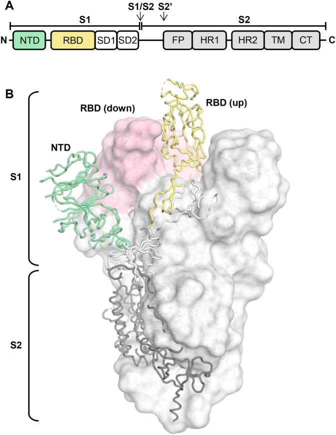
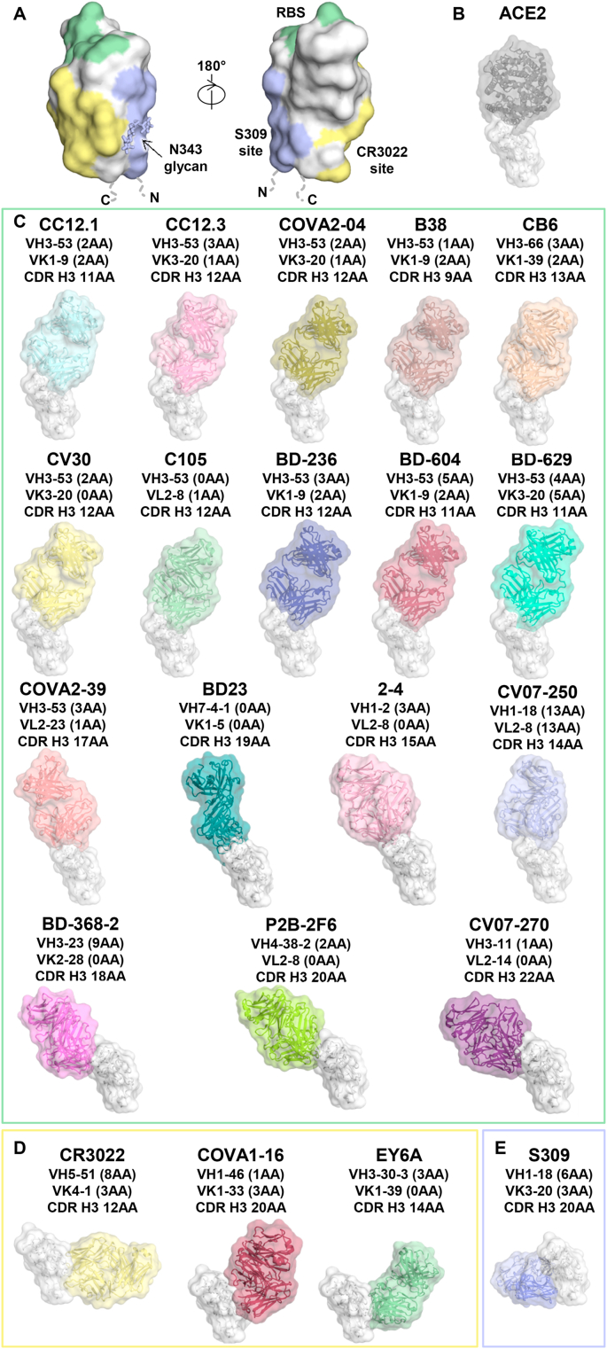
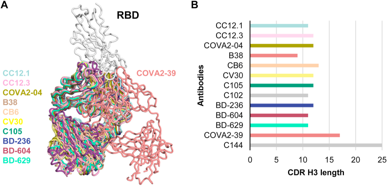
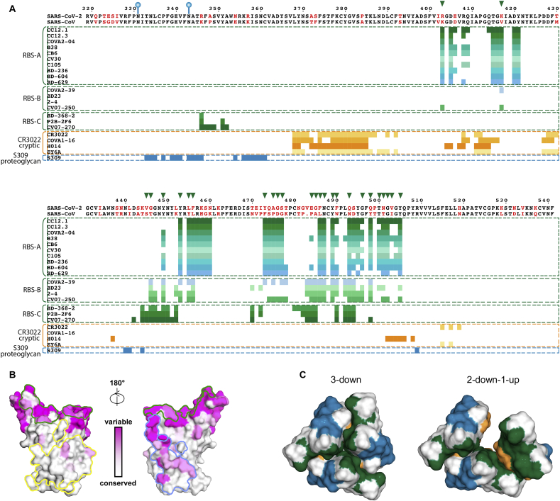
Immediately from the outset of the COVID-19 pandemic, researchers from diverse biomedical and biological disciplines have united to study the novel pandemic virus, SARS-CoV-2. The antibody response to SARS-CoV-2 has been a major focus of COVID-19 research due to its clinical relevance and importance in vaccine and therapeutic development. Isolation and characterization of antibodies to SARS-CoV-2 have been accumulating at an unprecedented pace. Most of the SARS-CoV-2 neutralizing antibodies to date target the spike (S) protein receptor binding domain (RBD), which engages the host receptor ACE2 for viral entry. Here we review the binding sites and molecular features of monoclonal antibodies that target the SARS-CoV-2 RBD, including a few that also cross-neutralize SARS-CoV.

Keywords: Antibody avidity; Cross-neutralization; Epitopes; Germline-encoded motifs; Neutralizing antibodies; RBD natural mutations; Receptor binding domain (RBD); SARS-CoV; SARS-CoV-2.


Conflict of interest statement

Declaration of competing interest The authors declare that they have no known competing financial interests or personal relationships that could have appeared to influence the work reported in this paper.

Figures

Fig. 1
Fig. 1
Schematic and structure of the SARS-CoV-2 spike (S) protein. (A) Schematic of the SARS-CoV-2 S protein. The receptor binding domain (RBD) is colored in yellow. NTD: N-terminal domain; SD: subdomain (1 and 2); FP, fusion peptide; HR1, heptad repeat 1; HR2, heptad repeat 2; TM, transmembrane domain; CT, cytoplasmic tail. Arrows denote protease cleavage sites. (B) Structure of the SARS-CoV-2 S protein in the prefusion conformation with one RBD in an up conformation and two RBDs in the down conformation (PDB ID: 6VSB) [46]. For the S1 domain, RBD (in an up conformation) and NTD are highlighted in yellow and green, respectively, with the remainder of S1 and S2 shown in white and grey. For clarity, only one protomer is represented in tubes and the other two subunits are represented by a white molecular surface with their RBDs in a down conformation in pink.
Fig. 2
Fig. 2
The distribution of IGHV gene usage of RBD-targeting neutralizing antibodies. A total of 280 antibodies reported in 19 studies are analyzed [[5], [6], [7], [8], [9], [10], [11], [12], [13], [14], [15], [16], [17], [18], [20], [21], [22], [23], [24]].
Fig. 3
Fig. 3
Antibody recognition of the RBD of SARS-CoV-2. (A) The main antigenic sites on the RBD of SARS-CoV-2 that have been identified to date. The RBD is shown in white, with the receptor-binding site (RBS) in green, CR3022 epitope in yellow, and S309 epitope in blue. (BE) Structures of the SARS-CoV-2 RBD with (B) human ACE2 receptor (PDB ID: 6M0J) [34], and (CE) neutralizing antibodies isolated from human patients. The RBDs (white) are all shown in the same relative orientation. Numbers of somatic mutated residues are shown in brackets after the light and heavy chain germline designation. The length of CDR H3 is indicated using the IMGT definition. (C) Structures of the RBD in complex with RBS-targeting antibodies: CC12.1 (PDB ID: 6XC2) [27], CC12.3 (PDB ID: 6XC4) [27], COVA2-04 (PDB ID: 7JMO) [28], B38 (PDB ID: 7BZ5) [23], CB6 (PDB ID: 7C01) [13], CV30 (PDB ID: 6XE1) [29], C105 (PDB ID: 6XCM) [30], BD-236 (PDB ID: 7CHB) [7], BD-604 (PDB ID: 7CH4) [7], BD-629 (PDB ID: 7CH5) [7], COVA2-39 (PDB ID: 7JMP) [28], BD23 (PDB ID: 7BYR) [16], 2–4 (PDB ID: 6XEY) [15], CV07-250 (PDB ID: 6XKQ) [6], BD-368-2 (PDB ID: 7CHE) [7], P2B-2F6 (PDB ID: 7BWJ) [17], and CV07-270 (PDB ID: 6XKP) [6]. (D) Structures of the RBD in complex with antibodies targeting the CR3022-binding site: CR3022 (PDB ID: 6W41) [31], COVA1-16 (PDB ID: 7JMW) [32], and EY6A (PDB ID: 6ZER) [14]. (E) Structure of antibody S309 with the RBD (PDB ID: 6WPT) [33].
Fig. 4
Fig. 4
Antibodies targeting the RBS-B epitope of SARS-CoV-2 RBD. Structures of four RBS-B antibodies are shown: BD23 (PDB ID: 7BYR) [16], COVA2-39 (PDB ID: 7JMP) [28], CV07-250 (PDB ID: 6XKQ) [6], and 2–4 (PDB ID: 6XEY) [15]. The RBD is shown in white, and the heavy and light chains of the bound antibodies are shown in dark and light colors, respectively. These antibodies approach the RBD at different angles and with different relative rotations of the Fab on the RBD surface. Interaction between Phe486 of the RBD and each antibody is shown in the right panels, with Phe486 highlighted in orange sticks.
Fig. 5
Fig. 5
Structural convergence of SARS-CoV-2 RBD-targeting antibodies encoded byIGHV3-53/366 genes. (A) Comparison of two completely different binding modes of antibodies encoded by IGHV3-53/3–66 germline genes that target the SARS-CoV-2 RBD and have different CDR H3 lengths [short (mode A) and long (mode B)]. All structures were retrieved from the PDB and superimposed on their RBD (white). (B) CDR H3 length (IMGT numbering) of the antibodies is shown in the bar chart. The color coding of the antibodies is the same as in Fig. 3. Structures of two other SARS-CoV-2 RBD-targeting antibodies encoded by IGHV3-53/3–66, namely C102 and C144 [35], are not publicly available and are not included in this figure.
Fig. 6
Fig. 6
Sequence conservation of the receptor-binding, CR3022, and S309 sites and epitope characterization. (A) Sequence alignment between RBDs of SARS-CoV-2 and SARS-CoV, with non-conserved residues highlighted in red. Residue numbers corresponding to SARS-CoV-2 RBD are labelled every ten residues above the sequence panel and the two N-glycosylation sites by blue balloons. Clusters of antibodies bound to different epitopes are shown between the colored dashed lines, with epitope residues represented as colored bars under the sequence alignment panel. Interactions between the RBD and its ligands including antibodies and ACE2 were analyzed using PISA program [83] with the same PDB accession codes as in Fig. 3 for RBS antibodies and 6W41, 7JMW, 7CAH, 6ZCZ, 6WPS, 6M0J for CR3022, COVA1-16, H104, EY6A, S309, ACE2 in complex with RBD, respectively. The analysis here uses buried surface (BSA > 0 Å2) as the criterion rather than contact residues for defining the epitope. Epitopes of RBS antibodies are shown in various shades of green and light blue, CR3022 cryptic site antibodies in various shades of yellow and orange, and S309 proteoglycan site antibody in dark blue. ACE2-binding residues are represented by green triangles on top of the sequence alignment. (B) Sequence conservation of the RBD [from white (high) to magenta (low)] among 17 SARS-like coronaviruses is highlighted on the RBD structure with the receptor-binding site outlined in green, CR3022 site in yellow, and S309 site in blue. (C) Spatial relationship among the epitope sites. To simplify the view, only epitopes of CC12.1 (green), COVA1-16 (orange) and S309 (blue) are selected to represent the RBD epitopes of the RBS, CR3022 cryptic site, and S309 proteoglycan site, respectively. Cryo-EM structures of the SARS-CoV-2 S protein (PDB ID 6VXX and 6VYB) are used to represent the 3-down and 2-down-1-up structures of the RBDs.
Fig. 7
Fig. 7
Natural variants of SARS-CoV-2 spike protein. Natural variants are highlighted in red, with sphere sizes corresponding to the frequency of each variant as reported in reference [72]. For clarity, only one protomer is represented in tubes and the other two are shown in a white surface (PDB ID: 6VSB4) [46]. (A) For the S1 domain, RBD (in an up conformation) and NTD are highlighted in yellow and green tubes, respectively, The S1 SD domains and S2 shown in light blue and grey, respectively. The rest of the S trimer is shown in a white surface representation. (B) Natural variants on the RBD are highlighted. (C) The major natural variant D614G is highlighted in red. D614 (red) can form a hydrogen bond with T859 (white) in an adjacent protomer, but G614 cannot form this H bond.

Similar articles

Cited by

References

    1. WHO Novel coronavirus – China. Jan 12, 2020. 2020. https://www.who.int/csr/don/12-january-2020-novel-coronavirus-china/en/
    1. Xu X., Chen P., Wang J., Feng J., Zhou H., Li X., Zhong W., Hao P. Evolution of the novel coronavirus from the ongoing Wuhan outbreak and modeling of its spike protein for risk of human transmission. Sci. China Life Sci. 2020;63:457–460. doi: 10.1007/s11427-020-1637-5. - DOI - PMC - PubMed
    1. Zhou P., Yang X.L., Wang X.G., Hu B., Zhang L., Zhang W., Si H.R., Zhu Y., Li B., Huang C.L., Chen H.D., Chen J., Luo Y., Guo H., Jiang R.D., Liu M.Q., Chen Y., Shen X.R., Wang X., Zheng X.S., Zhao K., Chen Q.J., Deng F., Liu L.L., Yan B., Zhan F.X., Wang Y.Y., Xiao G.F., Shi Z.L. A pneumonia outbreak associated with a new coronavirus of probable bat origin. Nature. 2020;579:270–273. doi: 10.1038/s41586-020-2012-7. - DOI - PMC - PubMed
    1. Premkumar L., Segovia-Chumbez B., Jadi R., Martinez D.R., Raut R., Markmann A., Cornaby C., Bartelt L., Weiss S., Park Y., Edwards C.E., Weimer E., Scherer E.M., Rouphael N., Edupuganti S., Weiskopf D., Tse L.V., Hou Y.J., Margolis D., Sette A., Collins M.H., Schmitz J., Baric R.S., de Silva A.M. The receptor binding domain of the viral spike protein is an immunodominant and highly specific target of antibodies in SARS-CoV-2 patients. Sci. Immunol. 2020;5 doi: 10.1126/sciimmunol.abc8413. - DOI - PMC - PubMed
    1. Han X., Wang Y., Li S., Hu C., Li T., Gu C., Wang K., Shen M., Wang J., Hu J., Wu R., Mu S., Gong F., Chen Q., Gao F., Huang J., Long Y., Luo F., Song S., Long S., Hao Y., Li L., Wu Y., Xu W., Cai X., Gao Q., Zhang G., He C., Deng K., Du L., Nai Y., Wang W., Xie Y., Qu D., Huang A., Tang N., Jin A. bioRxiv; 2020. A Rapid and Efficient Screening System for Neutralizing Antibodies and its Application for the Discovery of Potent Neutralizing Antibodies to SARS-CoV-2 S-RBD. - DOI - PMC - PubMed

Publication types

MeSH terms