Review
. 2021 Jan;191(1):52-65.
doi: 10.1016/j.ajpath.2020.10.001. Epub 2020 Oct 15.

Mechanisms of Endothelial Regeneration and Vascular Repair and Their Application to Regenerative Medicine

Affiliations
Review

Mechanisms of Endothelial Regeneration and Vascular Repair and Their Application to Regenerative Medicine

Colin E Evans et al. Am J Pathol. 2021 Jan.

Abstract

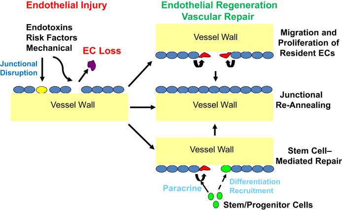
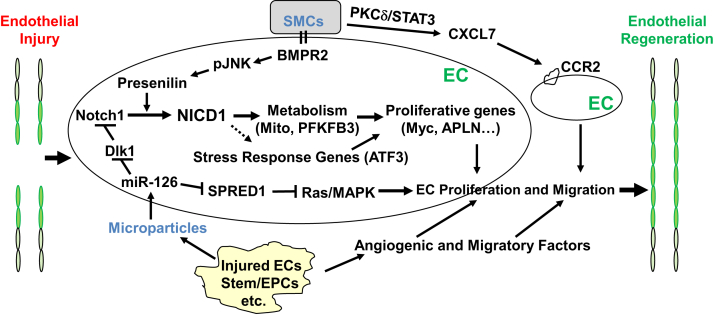
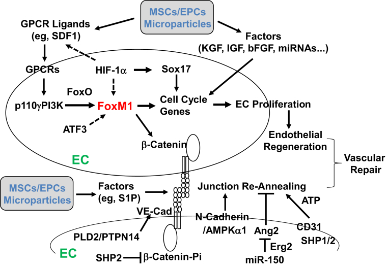
Endothelial barrier integrity is required for maintaining vascular homeostasis and fluid balance between the circulation and surrounding tissues and for preventing the development of vascular disease. Despite comprehensive understanding of the molecular mechanisms and signaling pathways that mediate endothelial injury, the regulatory mechanisms responsible for endothelial regeneration and vascular repair are incompletely understood and constitute an emerging area of research. Endogenous and exogenous reparative mechanisms serve to reverse vascular damage and restore endothelial barrier function through regeneration of a functional endothelium and re-engagement of endothelial junctions. In this review, mechanisms that contribute to endothelial regeneration and vascular repair are described. Targeting these mechanisms has the potential to improve outcome in diseases that are characterized by vascular injury, such as atherosclerosis, restenosis, peripheral vascular disease, sepsis, and acute respiratory distress syndrome. Future studies to further improve current understanding of the mechanisms that control endothelial regeneration and vascular repair are also highlighted.


Figures

Figure 1
Figure 1
Processes of endothelial regeneration and vascular repair. Endothelial injury induced by inflammatory or mechanical stimuli as well as risk factors are characterized by endothelial cell (EC) death and/or disruption of endothelial cell-cell junctions, leading to increases in vascular permeability. The vascular repair process involves restoration of a functional endothelial monolayer (ie, endothelial regeneration) and reannealing of the endothelial junctions to restore a semipermeable barrier. Endothelial regeneration is primarily attributable to migration and proliferation of resident ECs. Evidence of bone marrow–derived stem/progenitor cell engraftment is limited (dashed line), but these cells can contribute to endothelial regeneration in a paracrine manner through the release of regenerative/reparative factors.
Figure 2
Figure 2
Molecular and signaling mechanisms of endothelial regeneration and vascular repair in systemic arterial vessels after endothelial injury induced by mechanical or electrical denudation. Several studies that used carotid artery injury models have found an important role of endothelial Notch1 activation in the mechanism of endothelial proliferation. Microparticle-released miR-126 also plays an important role in endothelial proliferation and migration through inhibition of the Notch inhibitor delta-like 1 (Dlk1) and sprout-related EVH1 domain-containing protein 1 (SPRED1), Ras, and mitogen-activated protein kinase (MAPK) signaling. Smooth muscle cell (SMC) interaction with endothelial cells (ECs) promotes Notch1 activation through bone morphogenic protein receptor 2 (BMPR2) signaling, leading to EC proliferation. SMCs also release various factors (eg, CXCL7) to activate CCR2 signaling in the neighboring ECs and promote their migration for reendothelialization. Injured ECs and other cells, such as stem/progenitor cells, release microparticles and angiogenic and migratory factors and therefore promote endothelial regeneration. In an aorta mechanical pressure injury model, the stress response gene ATF3 plays an important role in mediating endothelial regeneration and vascular repair. NICD1, notch1 intracellular domain; PFKFB3, phosphofructo-2-kinase/fructose-2,6-biphosphatase 3; pJNK, phospho-c-Jun N-terminal kinase; PKC-δ, protein kinase Cδ.
Figure 3
Figure 3
Molecular and signaling mechanisms of endothelial regeneration and vascular repair in pulmonary vasculature after inflammatory injury. Studies have found a critical role of the transcriptional factor FoxM1 in mediating endothelial cell (EC) proliferation and endothelial regeneration after inflammatory vascular injury. FoxM1 is induced in lung vascular ECs only in the recovery phase after injury in a p110γ isoform of phosphoinositide 3-kinase (p110γPI3K)–dependent manner. Recent studies have further found that hypoxia-inducible factor (HIF)-1α mediates expression of FoxM1 and Sox17 to promote endothelial regeneration. Transplanted exogenous stem/progenitor cells act in a paracrine mechanism by releasing various growth factors, including stromal cell–derived factor 1 (SDF1) and insulin-like growth factor (IGF) as well as miRNA. Endothelial FoxM1 is one of the endogenous mediators of the paracrine effects. Vascular repair also involves reannealing of endothelial cell-cell junctions to reform a semipermeable barrier. One study found that β-catenin is the transcriptional target of FoxM1. Endothelial FoxM1 deficiency impairs endothelial barrier recovery though defective β-catenin expression. Other studies also found a role of Src homology 2 (SHP2) dephosphorylation of vascular endothelial cadherin (VE-cadherin)–associated β-catenin and phospholipase D2 (PLD2)/protein tyrosine phosphatase nonreceptor type 14 (PTPN14) dephosphorylation of VE-cadherin, AMP-activated protein kinase (AMPK)-α1/N-cadherin, and miR-150/Erg2/angiopoietin-2 (Ang2) signaling as well as stem/progenitor cell–released reparative factors, such as sphingosine-1 phosphate (S1P), in re-annealing the endothelial cell-cell junctions and restoring the endothelial barrier after injury. bFGF, basic fibroblast growth factor; EPCs, endothelial progenitor cells; GPCR, G-coupled protein receptor; KGF, keratinocyte growth factor; MSCs, mesenchymal stem cells.

References

    1. Deanfield J.E., Halcox J.P., Rabelink T.J. Endothelial function and dysfunction: testing and clinical relevance. Circulation. 2007;115:1285–1295. - PubMed
    1. Cines D.B., Pollak E.S., Buck C.A., Loscalzo J., Zimmerman G.A., McEver R.P., Pober J.S., Wick T.M., Konkle B.A., Schwartz B.S., Barnathan E.S., McCrae K.R., Hug B.A., Schmidt A.M., Stern D.M. Endothelial cells in physiology and in the pathophysiology of vascular disorders. Blood. 1998;91:3527–3561. - PubMed
    1. Libby P., Ridker P.M., Maseri A. Inflammation and atherosclerosis. Circulation. 2002;105:1135–1143. - PubMed
    1. Aird W.C. The role of the endothelium in severe sepsis and multiple organ dysfunction syndrome. Blood. 2003;101:3765–3777. - PubMed
    1. De Backer D., Creteur J., Preiser J.C., Dubois M.J., Vincent J.L. Microvascular blood flow is altered in patients with sepsis. Am J Respir Crit Care Med. 2002;166:98–104. - PubMed

Publication types

LinkOut - more resources