Review
. 2020 Dec:290:198197.
doi: 10.1016/j.virusres.2020.198197. Epub 2020 Oct 16.

The role of dysregulated immune responses in COVID-19 pathogenesis

Affiliations
Review

The role of dysregulated immune responses in COVID-19 pathogenesis

S Tahaghoghi-Hajghorbani et al. Virus Res. 2020 Dec.

Abstract

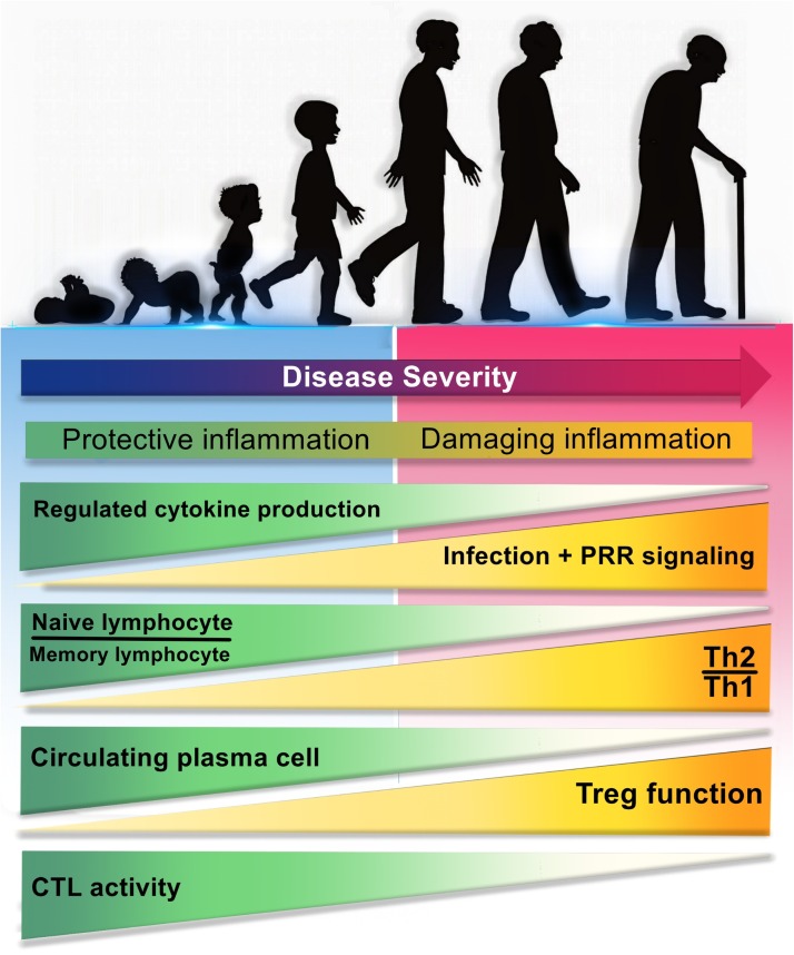
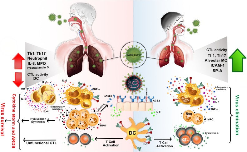
The coronavirus disease-2019 (COVID-19) which caused by severe acute respiratory syndrome-related coronavirus (SARS-CoV-2), is a pandemic threat to global public health. It has a wide spectrum of clinical manifestations from mild to critical illness, the most serious of which is the complications of acute respiratory distress syndrome (ARDS). SARS-CoV-2 infection appears mild in infants and children, however, in adults, it can lead to serious consequences. In this review, we highlighted the differences between the immune responses of the lung in children and adults, immune dysregulation and their possible role in clinical manifestations in COVID-19. There is a reduction in population of immunocompetent cells during aging and subsequently induced ineffective inflammation in the faces of some infections. Dysregulation in the immune system can lead to an unappropriated local and systemic immune responses and subsequently the rapid spread of the virus, leading to severe COVID-19 disease. Therefore, recognizing the differences in the immune responses of various hosts as well as to improve the immune system disorder should always be part of research and treatment protocols.

Keywords: COVID-19; Immune dysregulation; Immunopathogenesis; SARS-CoV-2.


Conflict of interest statement

The authors declare no conflicts of interests

Figures

Fig. 1
Fig. 1
The relationship between age-dependent changes in immune responses with the severity of COVID-19 disease. Differences in the immune system of children and adults may be the reasons for clinical differences in the severity of COVID-19. During aging, immune responses undergo changes that lead to more severe disease, some of which are include: a) depletion of well-ordinated innate immunity and regulative cytokines, b) diminished ability of the innate immune cells to recognize PAMPs, followed by strong activation of PRRs, influx of pathogenic immune cells and excessive release of proinflammatory cytokines for compensation, c) reduction ratio of naïve lymphocyte/memory lymphocyte, d) induce of negative regulation and the predominance of Th2 to Th1 responses, e) decrement of circulating plasma cells, f) increase of regulatory T cells (CD4+ CD25+ FOXP3+) function, and g) decrease of CTLs activity and diminish of CTL-mediated immunity. PAMP: Pathogen associated molecular pattern; PRR: Pathogen recognition receptor; Th: T helper; CTL: Cytotoxic T lymphocyte.
Fig. 2
Fig. 2
The difference in immune responses in the lungs of children and adults to SARS-CoV-2 is the reason for the different clinical manifestations. In children, SARS−COV-2 infection may be quickly eradicated due to having less mature ACE2 receptors and rapid activation of immunocompetent immune cells (right side). In adults, the negative regulation of the immune response in the respiratory tract, late changes in the nature of the immune responses, decrease in population of immunocompetent cells, increase of ACE2 expression, ACE2 shedding and sACE2 production, all can lead to an uncontrolled immune response, widespread ineffective inflammation, immune dysregulation, cytokine storm and ARDS (left side). ACE2: Angiotensin−COnverting enzyme 2; SARS−COV2: severe acute respiratory syndrome coronavirus 2; ARDS: Acute respiratory distress syndrome.
Fig. 3
Fig. 3
SARS-CoV-2 biogenesis cycle and probably action site of commonly used drugs in the early stage of acute infection. TMPRSS2 cleaves and activates SARS−COV-2 -S glycoprotein for binding to its target receptor (ACE2) on the epithelial cells of the respiratory tracts. After receptor binding, the virus enters, gains access to the host cell cytosol and replicates. Following subgenomic RNA synthesis and assembly, virions are transported to the cell surface in vesicles and released by exocytosis. There are some potential approaches to prevent virus entry to the target cells and blocks several stages of life cycle of SARS−COV-2, for instance attachment, clathrin-mediated endocytosis, and replication. 1a > Fusion, 1b > Endocytosis, 2> Viral-host membrane fusion and release of viral RNA, 3> Translation, 4> Proteolysis, 5> Trafficking of newly synthesized viral proteins to the Golgi, 6> Transcription and replication of viral RNA, 7a > Assembly of mature Virion in a vesicle, 7b > Final Packaging, 8> Virion release via exocytosis. ACE2: Angiotensin−COnverting enzyme 2; SARS−COV2: severe acute respiratory syndrome coronavirus2.

References

    1. Beniac D.R., Andonov A., Grudeski E., Booth T.F. Architecture of the SARS coronavirus prefusion spike. Nat. Struct. Mol. Biol. 2006;13(8):751–752. - PMC - PubMed
    1. Delmas B., Laude H. Assembly of coronavirus spike protein into trimers and its role in epitope expression. J. Virol. 1990;64(11):5367–5375. - PMC - PubMed
    1. Li G., Fan Y., Lai Y., Han T., Li Z., Zhou P. Coronavirus infections and immune responses. J. Med. Virol. 2020;92(4):424–432. - PMC - PubMed
    1. Raj V.S., Mou H., Smits S.L., Dekkers D.H., Müller M.A., Dijkman R. Dipeptidyl peptidase 4 is a functional receptor for the emerging human coronavirus-EMC. Nature. 2013;495(7440):251–254. - PMC - PubMed
    1. Pene F., Merlat A., Vabret A., Rozenberg F., Buzyn A., Dreyfus F. Coronavirus 229E-related pneumonia in immunocompromised patients. Clin. Infect. Dis. 2003;37(7):929–932. - PMC - PubMed