. 2020 Dec;472(12):1705-1717.
doi: 10.1007/s00424-020-02469-z. Epub 2020 Oct 17.

Neurogenic tachykinin mechanisms in experimental nephritis of rats

Affiliations

Neurogenic tachykinin mechanisms in experimental nephritis of rats

Kristina Rodionova et al. Pflugers Arch. 2020 Dec.

Abstract

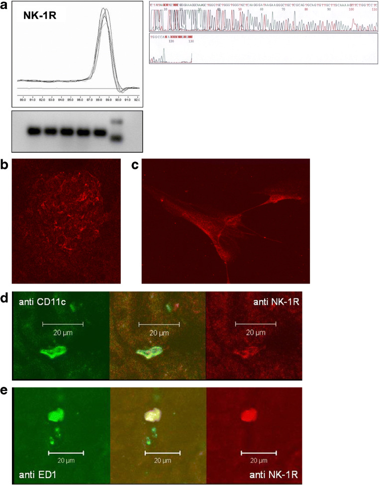
We demonstrated earlier that renal afferent pathways combine very likely "classical" neural signal transduction to the central nervous system and a substance P (SP)-dependent mechanism to control sympathetic activity. SP content of afferent sensory neurons is known to mediate neurogenic inflammation upon release. We tested the hypothesis that alterations in SP-dependent mechanisms of renal innervation contribute to experimental nephritis. Nephritis was induced by OX-7 antibodies in rats, 6 days later instrumented for recording of blood pressure (BP), heart rate (HR), drug administration, and intrarenal administration (IRA) of the TRPV1 agonist capsaicin to stimulate afferent renal nerve pathways containing SP and electrodes for renal sympathetic nerve activity (RSNA). The presence of the SP receptor NK-1 on renal immune cells was assessed by FACS. IRA capsaicin decreased RSNA from 62.4 ± 5.1 to 21.6 ± 1.5 mV s (*p < 0.05) in controls, a response impaired in nephritis. Suppressed RSNA transiently but completely recovered after systemic administration of a neurokinin 1 (NK1-R) blocker. NK-1 receptors occurred mainly on CD11+ dendritic cells (DCs). An enhanced frequency of CD11c+NK1R+ cell, NK-1 receptor+ macrophages, and DCs was assessed in nephritis. Administration of the NK-1R antagonist aprepitant during nephritis reduced CD11c+NK1R+ cells, macrophage infiltration, renal expression of chemokines, and markers of sclerosis. Hence, SP promoted renal inflammation by weakening sympathoinhibitory mechanisms, while at the same time, substance SP released intrarenally from afferent nerve fibers aggravated immunological processes i.e. by the recruitment of DCs.

Keywords: Dendritic cells; Nephritis; Renal innervation; Sympathetic nerve activity; Tachykinins.


Figures

Fig. 1
Fig. 1
Decreases of renal sympathetic nerve activity after four administrations of a 10 μl bolus of capsaicin or saline into the kidney (filled circles—healthy animals; open triangle—rats with anti-Thy1.1 nephritis; open circle—control group injected with saline). In healthy rats, the capsaicin administration resulted in a steep decrease in sympathetic nerve activity, which was highly significantly reduced in animals with nephritis. Intrarenal saline administration had no effect. Intravenous administration of the substance P receptor antagonist RP67580 (10 mg/kg) was followed by short steep increases in sympathetic nerve activity above baseline levels (duration less than 1 min) in nephritic rats as well as in control animals suggesting SP release from afferent renal nerve fibers upon intrarenal TRPV1 receptors by capsaicin. Interestingly, baseline activity also increased significantly in saline-treated rats after administration of RP67580. This indicates that a tonic SP-dependent sympathoinhibition must also be effective under resting conditions (n = 6; #*p < 0.05 versus baseline)
Fig. 2
Fig. 2
a Renal cortical NK-1R mRNA expression of untreated rats was detected by real-time RT-PCR. Amplified PCR products of NK-1R showed identical and specific melting curves as well as an identical and expected length of 125 base pairs in agarose gel electrophoresis. Subsequent sequence analysis revealed a 97% congruence to the published mRNA rat NK-1R transcript. Immunofluorescence analysis by confocal laser scanning microscopy showed NK-1R (red) in a glomerulus (b), on cultured mesangial cells (c), on ED1-positive macrophages (green; d), and on CD11c-positive dendritic cells (green; e). The yellow merge indicates colocalization of NK-1R with ED1-positive cells and CD11c-positive dendritic cells, respectively
Fig. 3
Fig. 3
NK1-R expression on macrophages in glomerulonephritis. Rats with anti-Thy1.1-induced glomerulonephritis (filled bars) or with saline (stripped bar) for control group. Frequency of macrophages (a), ED1+NK1R+ cells among lymphocytes (b) as well as ED1+NK1R+ cells among macrophages (c), and the mean fluorescence intensity of NK-1R expression on macrophages (d) were detected by FASC analysis. Data are given as mean ± SEM; n = 6, *p < 0.05 versus saline; #p < 0.05 versus saline
Fig. 4
Fig. 4
NK1-R expression on T cells in glomerulonephritis. Rats with anti-Thy1.1-induced antibody glomerulonephritis (filled bars) or with saline (stripped bar) for control group. NK1-R expression was measured by FACS analysis. Although the frequency of T cells is increased (a), only a small percentage of CD3-positive T cells express NK-1R (b). Data are given as mean ± SEM; n = 6, *p < 0.05 versus saline; #p < 0.05 versus saline
Fig. 5
Fig. 5
NK1-R expression on dendritic cells in glomerulonephritis. Rats with anti-Thy1.1 antibody induced glomerulonephritis (filled bars) or with saline (stripped bar) as control group. NK1-R expression was measured by FACS analysis. Frequency of NK-1R+CD11c+ among lymphocytes (a) as well as among dendritic cells (b) was increased 48 h after antibody induction (b). Data are given as mean ± SEM; *p < 0.05 versus saline; #p < 0.05 versus 6 days (6d)
Fig. 6
Fig. 6
Aprepitant exerts an anti-inflammatory effect in anti-Thy1.1 nephritis. Rats were pretreated with 5 mg/kg aprepitant i.p. 12 h and 1 h before injection of anti-Thy1.1 mAb and every 24 h after antibody challenge (diagonal bars). Further animals received vehicle plus anti-Thy1.1 (black bars) or aprepitant plus saline instead of anti-Thy1.1 mAb (white bars). Renal cortical mRNA expression of TNFα, PAI-1, IL-6, and RANTES was detected by real-time RT-PCR. The relative amount of mRNA was normalized against the housekeeping gene β-actin. Data are given as mean ± SEM; *p < 0.05 versus aprepitant plus vehicle; #p < 0.05 versus anti Thy1.1 mAb plus aprepitant
Fig. 7
Fig. 7
NK-1R blockade decreases protein synthesis of TNFα. Rats were pretreated with 5 mg/kg aprepitant i.p. 12 h and 1 h before injection of anti-Thy1.1 mAb and every 24 h after antibody challenge. TNFα protein was detected by Western Blot and GAPDH was used as house keeping gene to ensure equally protein amounts. The small western blot shows pooled lysates of renal cortical tissues of all three groups (a). b Saline plus anti-Thy-1.1 mAb and aprepitant plus anti-Thy1.1 mAb were quantified. Data are given as mean ± SEM; n = 6; *p < 0.05
Fig. 8
Fig. 8
Aprepitant pretreatment reduced renal macrophages (a and b), TGFβ expression (c), deposition of collagen IV (d), and interstitial proliferation (e) anti-Thy1.1 nephritis. Rats were pretreated with 5 mg/kg aprepitant i.p. 12 h and 1 h before injection of anti-Thy1.1 mAb and every 24 h after antibody challenge (diagonal bars). Control animals received either vehicle plus anti-Thy1.1 mAb (black bars) or aprepitant plus saline instead of anti-Thy1.1 mAb (white bars). Alterations of macrophages were determinated by immunohistochemistry. Renal cortical mRNA expression of TGFβ was detected by real-time RT-PCR. The relative amount of mRNA was normalized against the housekeeping gene β-actin. Data are given as mean ± SEM; n = 6, *p < 0.05 versus aprepitant plus vehicle; #p < 0.05 versus anti Thy 1.1 mAb plus aprepitant

References

    1. Abdulla MH, Johns EJ. The innervation of the kidney in renal injury and inflammation: a cause and consequence of deranged cardiovascular control. Acta Physiol (Oxford) 2017;220:404–416. doi: 10.1111/apha.12856. - DOI - PubMed
    1. Ascon D, Lopez-Briones S, Liu M, Ascon M, Savransky V, Colvin R, Soloski M, Rabb H. Phenotypic and functional characterization of kidney-infiltrating lymphocytes in renal ischemia reperfusion injury. J Immunol. 2006;177:3380–3387. doi: 10.4049/jimmunol.177.5.3380. - DOI - PubMed
    1. Bang R, Sass G, Kiemer AK, Vollmar AM, Neuhuber WL, Tiegs G. Neurokinin-1 receptor antagonists CP-96,345 and L-733,060 protect mice from cytokine-mediated liver injury 6. J Pharmacol ExpTher. 2003;305:31–39. doi: 10.1124/jpet.102.043539. - DOI - PubMed
    1. Bernard C (1859) Leçons sur les Propriétés et les Altérations Pathologiques des Liquides de L’Organisme. Bailliére et Fils, Paris: vol 2, 170
    1. Bhatt DL, Kandzari DE, O'Neill WW, D'Agostino R, Flack JM, Katzen BT, Leon MB, Liu M, Mauri L, Negoita M, Cohen SA, Oparil S, Rocha-Singh K, Townsend RR, Bakris GL. A controlled trial of renal denervation for resistant hypertension. N Engl J Med. 2014;370:1393–1401. doi: 10.1056/NEJMoa1402670. - DOI - PubMed

Publication types

MeSH terms