. 2021 Mar;238(3):527-535.
doi: 10.1111/joa.13335. Epub 2020 Oct 18.

Ultrastructural evidence for telocytes in equine tendon

Affiliations

Ultrastructural evidence for telocytes in equine tendon

María J Luesma et al. J Anat. 2021 Mar.

Abstract

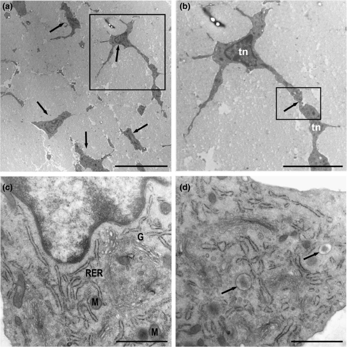
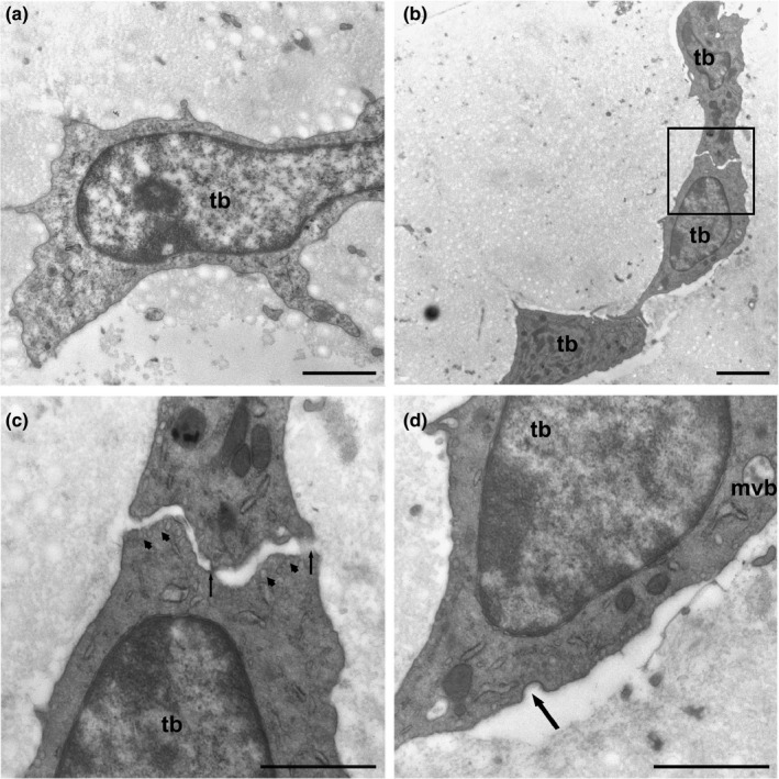
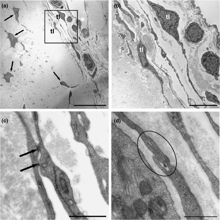
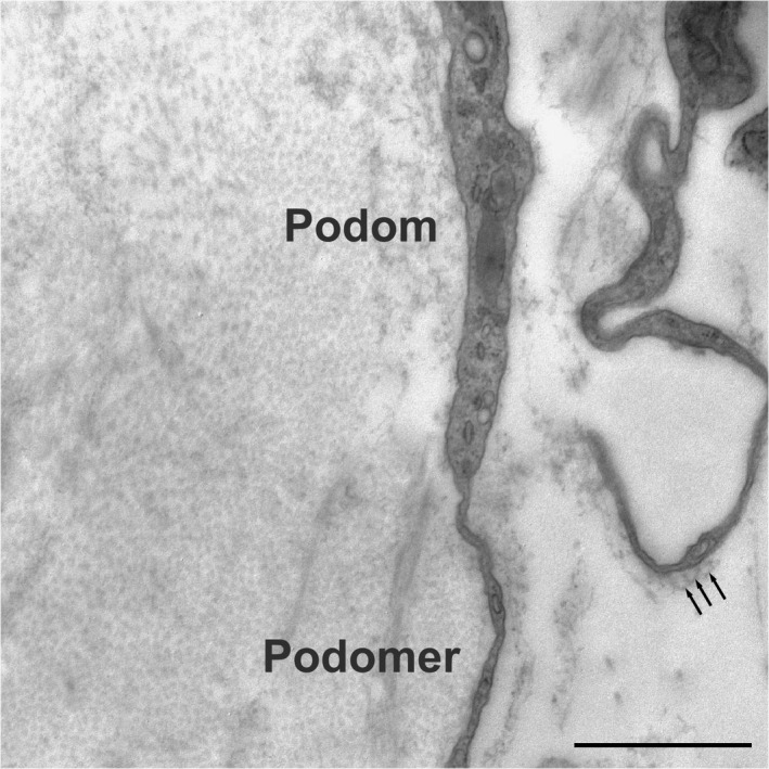
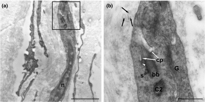
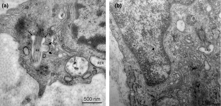
The three-dimensional ultrastructure of the tendon is complex. Two main cell types are classically supported: elongated tenocytes and ovoid tenoblasts. The existence of resident stem/progenitor cells in human and equine tendons has been demonstrated, but their location and relationship to tenoblasts and tenocytes remain unclear. Hence, in this work, we carried out an ultrastructural study of the equine superficial digital flexor tendon. Although the fine structure of tendons has been previously studied using electron microscopy, the presence of telocytes, a specific type of interstitial cell, has not been described thus far. We show the presence of telocytes in the equine inter-fascicular tendon matrix near blood vessels. These telocytes have characteristic telopodes, which are composed of alternating dilated portions (podoms) and thin segments (podomers). Additionally, we demonstrate the presence of the primary cilium in telocytes and its ability to release exosomes. The location of telocytes is similar to that of tendon stem cells. The telocyte-blood vessel proximity, the presence of primary immotile cilia and the release of exosomes could have special significance for tendon homeostasis.

Keywords: primary cilium; telocytes; tendon; tenocytes; transmission electron microscopy.


Conflict of interest statement

The authors confirm that there are no conflicts of interest.

Figures

FIGURE 1
FIGURE 1
Ultrathin sections of the equine digital flexor tendon. (a) Tendon areas showing polygonal and stellate tenocytes (arrows) immersed in a wide area of extracellular matrix. (b) Detail of A‐Tenocytes (Tn) with long slender cytoplasmic processes that contact neighbouring tenocytes (inset). (c) Tenocytes with rough endoplasmic reticulum (RER), a well‐developed Golgi apparatus (G) and few mitochondria (M). The nucleus contains a narrow layer of marginal chromatin. (d) A large number of vesicles released from the Golgi apparatus showing an electron‐dense central core containing matrix proteins were observed (arrows). Scale bars: (a) 10 µm; (b) 5 µm; (c) 1 µm; (d) 1 µm
FIGURE 2
FIGURE 2
(a) Tenoblast (tb) showing a voluminous nucleus and prominent nucleolus. (b) The cytoplasmic prolongations were wider and shorter than those in tenocytes and established contacts with neighbouring tenoblasts (inset). (c) Detail of B‐Tenoblast membranes made punctate contacts with electron‐dense reinforcements (arrows). Numerous pinocytic vesicles appear below the plasma membrane (arrowheads). (d) Tenoblasts (tb) have small saccules of rough endoplasmic reticulum and multivesicular bodies (mvb) containing exosomes. Numerous pinocytic vesicles and coated pits are indicative of endocytic processes mediated by clathrin (arrow). Scale bars: (a) 1 µm; (b) 2 µm; (c) 1 µm; (d) 1 µm
FIGURE 3
FIGURE 3
(a) Tenocytes (arrows) embedded in the extracellular matrix. Various telocytes (tl) can be observed around the tendon capillaries (inset). (b) Telocytes (tl) showed voluminous nuclei and scant perinuclear cytoplasm (c) and extended via very long extensions to make contact with neighbouring cells through gap‐like junctions (arrows), and (d) peg‐and‐socket contacts (encircled). Scale bars: (a) 20 µm; (b) 5 µm; (c) 1 µm; (d) 0.5 µm
FIGURE 4
FIGURE 4
The prolongations of the telocytes, named telopodes, comprised dilated portions named podoms containing mitochondria, small saccules of RER, and filiform segments named podomers. The podomers were observed in relation to small vesicles (exosomes) (arrows). Scale bar: 1 µm
FIGURE 5
FIGURE 5
(a) Location of the primary cilium of a telocyte (inset). n: nucleus. (b) Detail of A‐Primary cilium emerging from a basal body (bb). The proximal region of the cilium is located within an invagination of the plasma membrane (ciliary pocket) (white arrows) (cp). The distal end of the basal body (bb) contacts the plasma membrane through transitional fibres (arrowheads). Extracellular vesicles can be observed in close relationship with the primary cilium (black arrows). C2, daughter centriole; G, Golgi apparatus; S, satellite. Scale bars: (a) 2 µm; (b) 0.5 µm
FIGURE 6
FIGURE 6
Pseudoglandular, canalicular, and saccular phase of lung development (a‐c). Exemplary micro‐CT images of coronary cross sections at ED15 (a), ED20 (b), and N0 (c). Additional histological sections (H&E) of corresponding pulmonary tissue (×10 magnification; d,e,f). Images were adjusted for best visualization. In the pseudoglandular stage, lung parenchyma had a glandular structure and terminal buds grew out into the surrounding mesenchyme (a,d). In the later stages, branching and penetration of the epithelial tubules into the surrounding tissue were observed. During the canalicular stage the airways expanded (b,e). In the saccular stage, transition from branching of the airways to alveolarization could be observed (c,f). Scalebar 1 mm

References

    1. Aleksandrovych, V. , Pasternak, A. , Basta, P. , Sajewicz, M. , Walocha, J.A. & Gil, K. (2017) Telocytes: facts, speculations and myths (Review article). Folia Medica Cracoviensia, 57(1), 5–22. Available from: http://www.ncbi.nlm.nih.gov/pubmed/28608858 [Accessed: 12 December 2019] - PubMed
    1. Anvarian, Z. , Mykytyn, K. , Mukhopadhyay, S. , Pedersen, L.B. & Christensen, S.T. (2019) Cellular signalling by primary cilia in development, organ function and disease. Nature Reviews Nephrology, 15(4), 199–219. 10.1038/s41581-019-0116-9 - DOI - PMC - PubMed
    1. Bei, Y. , Wang, F. , Yang, C. & Xiao, J. (2015) Telocytes in regenerative medicine. Journal of Cellular and Molecular Medicine, 19(7), 1441–1454. 10.1111/jcmm.12594 - DOI - PMC - PubMed
    1. Benjamin, M. & Ralphs, J.R. (1997) Tendons and ligaments — an overview. Histology and Histopathology, 12(4), 1135–1144. - PubMed
    1. Bi, Y. , Ehirchiou, D. , Kilts, T.M. , Inkson, C.A. , Embree, M.C. , Sonoyama, W. et al. (2007) Identification of tendon stem/progenitor cells and the role of the extracellular matrix in their niche. Nature Medicine, 13(10), 1219–1227. 10.1038/nm1630 - DOI - PubMed

LinkOut - more resources