

Extra-pelvic endometriosis: A review
- PMID: 33071634
- PMCID: PMC7542014
- DOI: 10.1002/rmb2.12340
Extra-pelvic endometriosis: A review
Abstract
Background: Extra-pelvic endometriosis is a rare type of endometriosis, which occurs in a distant site from gynecological organs. The diagnosis of extra-pelvic endometriosis can be extremely challenging and may result in a delay in diagnosis. The main objective of this review was to characterize abdominal wall endometriosis (AWE) and thoracic endometriosis (TE).
Methods: The authors performed a literature search to provide an overview of AWE and TE, which are the major types of extra-pelvic endometriosis.
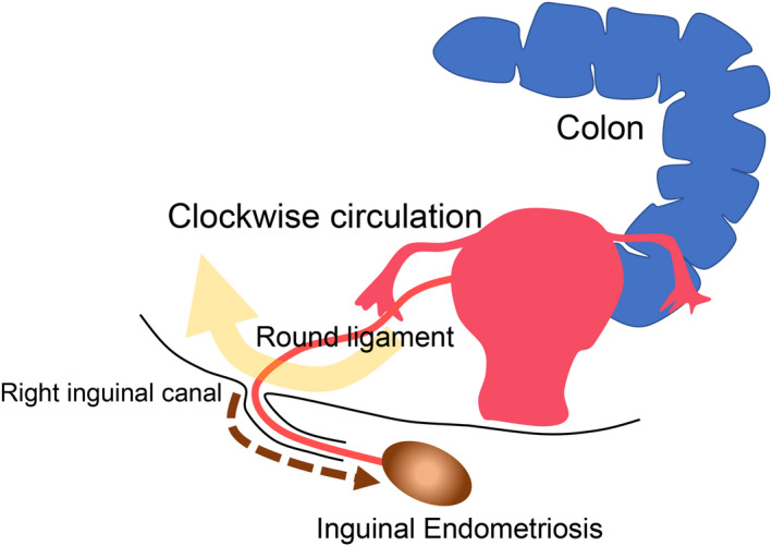
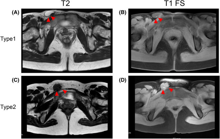
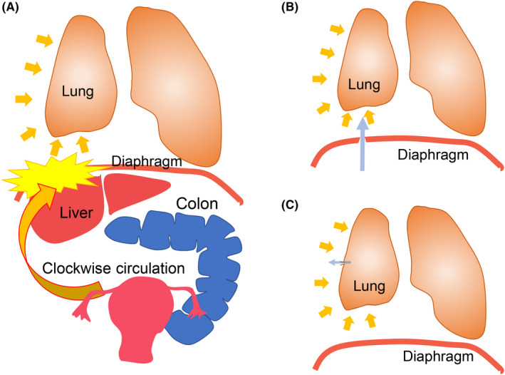
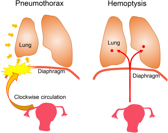
Main findings: Abdominal wall endometriosis includes scar endometriosis secondary to the surgical wound and spontaneous AWE, most of which occur in the umbilicus or groin. Surgical treatment appeared to be effective for AWE. Case reports indicated that the diagnosis and treatment of catamenial pneumothorax or endometriosis-related pneumothorax (CP/ERP) are challenging, and a combination of surgery and postoperative hormonal therapy is essential. Further, catamenial hemoptysis (CH) can be adequately managed by hormonal treatment, unlike CP/ERP.
Conclusion: Evidence-based approaches to diagnosis and treatment of extra-pelvic endometriosis remain immature given the low prevalence and limited quality of research available in the literature. To gain a better understanding of extra-pelvic endometriosis, it would be advisable to develop a registry involving a multidisciplinary collaboration with gynecologists, general surgeons, and thoracic surgeons.
Keywords: abdominal wall endometriosis; catamenial hemoptysis; catamenial pneumothorax; extra‐pelvic endometriosis; treatment.
© 2020 The Authors. Reproductive Medicine and Biology published by John Wiley & Sons Australia, Ltd on behalf of Japan Society for Reproductive Medicine.
Conflict of interest statement
Conflict of interest: The authors report no conflict of interest. Human rights statements and informed consent/Animal studies: This article does not contain any studies with human and animal subjects performed by any of the authors.
Figures
References
-
- Zondervan KT, Becker CM, Koga K, Missmer SA, Taylor RN, Vigano P. Endometriosis. Nat Rev Dis Primers. 2018;4(1):9. - PubMed
-
- Vercellini P, Vigano P, Somigliana E, Fedele L. Endometriosis: pathogenesis and treatment. Nat Rev Endocrinol. 2014;10(5):261‐275. - PubMed
-
- Sampson JA. Peritoneal endometriosis due to the menstrual dissemination of endometrial tissue into the peritoneal cavity. Am J Obstet Gynecol. 1927;14(4):422‐469.
-
- Giudice LC, Kao LC. Endometriosis. Lancet. 2004;364(9447):1789‐1799. - PubMed
LinkOut - more resources
Full Text Sources
Other Literature Sources
Miscellaneous