Review
. 2020 Sep 23:14:525970.
doi: 10.3389/fnins.2020.525970. eCollection 2020.

Local Sleep and Alzheimer's Disease Pathophysiology

Affiliations
Review

Local Sleep and Alzheimer's Disease Pathophysiology

Bryce A Mander. Front Neurosci. .

Abstract

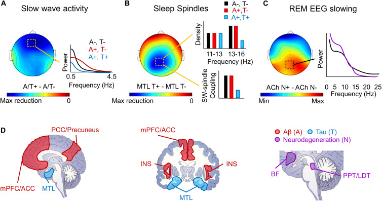
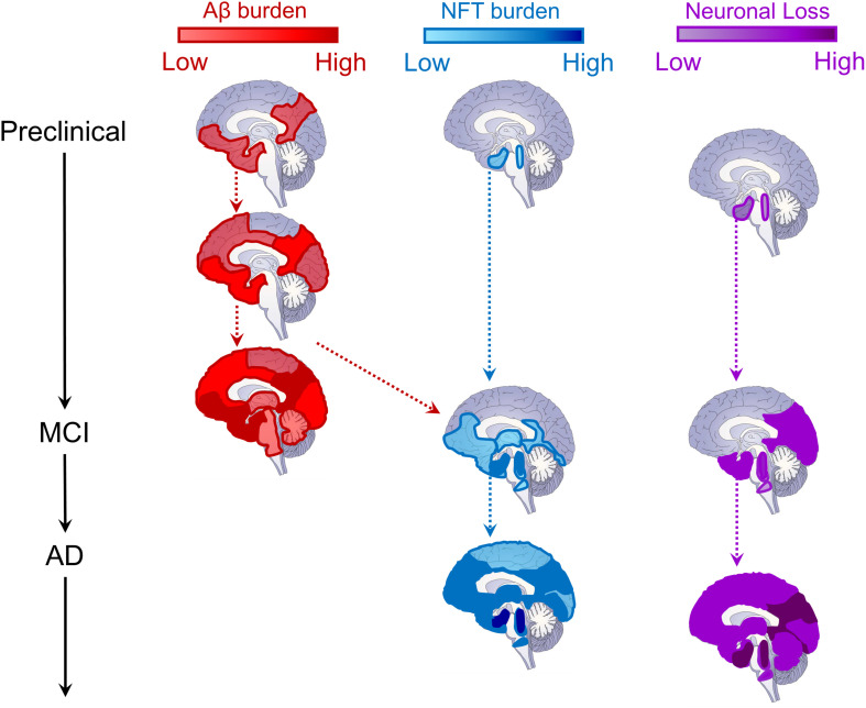
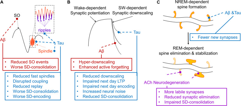
Even prior to the onset of the prodromal stages of Alzheimer's disease (AD), a constellation of sleep disturbances are apparent. A series of epidemiological studies indicate that multiple forms of these sleep disturbances are associated with increased risk for developing mild cognitive impairment (MCI) and AD, even triggering disease onset at an earlier age. Through the combination of causal manipulation studies in humans and rodents, as well as targeted examination of sleep disturbance with respect to AD biomarkers, mechanisms linking sleep disturbance to AD are beginning to emerge. In this review, we explore recent evidence linking local deficits in brain oscillatory function during sleep with local AD pathological burden and circuit-level dysfunction and degeneration. In short, three deficits in the local expression of sleep oscillations have been identified in relation to AD pathophysiology: (1) frequency-specific frontal deficits in slow wave expression during non-rapid eye movement (NREM) sleep, (2) deficits in parietal sleep spindle expression, and (3) deficits in the quality of electroencephalographic (EEG) desynchrony characteristic of REM sleep. These deficits are noteworthy since they differ from that seen in normal aging, indicating the potential presence of an abnormal aging process. How each of these are associated with β-amyloid (Aβ) and tau pathology, as well as neurodegeneration of circuits sensitive to AD pathophysiology, are examined in the present review, with a focus on the role of dysfunction within fronto-hippocampal and subcortical sleep-wake circuits. It is hypothesized that each of these local sleep deficits arise from distinct network-specific dysfunctions driven by regionally-specific accumulation of AD pathologies, as well as their associated neurodegeneration. Overall, the evolution of these local sleep deficits offer unique windows into the circuit-specific progression of distinct AD pathophysiological processes prior to AD onset, as well as their impact on brain function. This includes the potential erosion of sleep-dependent memory mechanisms, which may contribute to memory decline in AD. This review closes with a discussion of the remaining critical knowledge gaps and implications of this work for future mechanistic studies and studies implementing sleep-based treatment interventions.

Keywords: Alzheimer’s disease; REM sleep; beta-amyloid protein; neurodegeneration; sleep; sleep spindle; slow waves; tau and phospho-tau protein.


Figures

FIGURE 1
FIGURE 1
Schematic depiction of local sleep features disrupted by AD pathophysiology. (A) A schematic topoplot of decreases in SWA during NREM sleep in individuals with Aβ (A +, red) and tau (T +, blue) pathology relative to those without pathology (A–, T–, black) is presented, with cooler colors reflecting the severity in the hypothesized reduction in SWA. A schematic of the effects of Aβ and tau pathology on SWA by frequency are plotted to the right. (B) A topoplot of decreases in sleep spindles in individuals with tau pathology, particularly in the medial temporal lobe (MTL) is presented, with cooler colors reflecting the severity of sleep spindle loss in individuals with tau pathology. Schematic bar graphs of the effects of both Aβ (red) and tau (blue) pathology on sleep spindle density and slow wave (SW)-sleep spindle coupling relative to those without AD pathology (black) are presented to the right. (C) A topoplot of the increase in REM sleep EEG slowing in those with cholinergic degeneration (ACh N +, purple) relative to those without significant cholinergic degeneration (ACh N–, black). A schematic frequency by spectral power plot of REM sleep is shown to the right, indicating increased low frequency power and decreased high frequency power in the presence of cholinergic degeneration. (D) Schematic of brain slices depicting the hypothesized locations of Aβ (A, red) and tau (T, blue) deposition, as well as neurodegeneration (N, purple), that lead to the observed local deficits in NREM and REM sleep. mPFC/ACC denotes medial prefrontal cortex and anterior cingulate cortex, PCC/Precuneus denotes posterior cingulate cortex and precuneus, MTL denotes the medial temporal lobe, INS denotes the insula cortex, BF denotes the cholinergic basal forebrain, and PPT/LDT denotes the cholinergic pedunculopontine and lateral dorsal tegmental nuclei.
FIGURE 2
FIGURE 2
Schematic depiction of the evolution of Alzheimer’s disease biomarkers. Sagittal brain slices depicting the hypothesized locations of Aβ (red, left column) and tau (blue, middle column) deposition, as well as neurodegeneration (neuronal loss, purple, right column) across disease stages. For Aβ, deposition begins in medial prefrontal, cingulate, temporal, and precuneus regions, and progresses cortically and then subcortically until most of the brain contains amyloid plaques (Braak and Braak, 1991; Thal et al., 2002; Sepulcre et al., 2013; Palmqvist et al., 2017). The cortex is likely already saturated with amyloid plaques by the time most patients convert to MCI (Jack et al., 2010, 2013). For tau neurofibrillary tangles (NFT), deposition begins in the locus coeruleus and cholinergic brainstem nuclei in Braak stage 0, progressing into entorhinal cortex in the medial temporal lobe by stage I (Braak and Braak, 1991; Mesulam et al., 2004; Grudzien et al., 2007; Ehrenberg et al., 2017). NFTs remains largely constrained within the MTL until Braak stage III (Braak and Braak, 1991, 1995), when, through its interaction with Aβ pathology (red dashed arrow), tau pathology spreads throughout the brainstem, thalamus, MTL, inferior temporal cortex, and medial prefrontal and cingulate brain regions (Vogel et al., 2020). It is at this time that MCI conversion begins to be observed (Jack et al., 2010, 2013). In later Braak stages, tau pathology is observed throughout most of the cortex (Braak and Braak, 1991, 1995), triggering widespread cortical degeneration as its spreads (Jack et al., 2010, 2013). Regarding neurodegeneration (neuronal loss), as with NFTs, synaptic and neuronal loss begins within locus coeruleus, cholinergic brainstem nuclei, and the MTL (Mesulam et al., 2004; Grudzien et al., 2007; Whitwell, 2010). As MCI progresses into later stages, cortical atrophy is prevalent throughout the temporal, parietal, and occipital lobes (Whitwell, 2010). Ultimately, in later AD stages, cortical atrophy progresses through lateral frontal cortex and, eventually, throughout the entire brain, impacting neural regulation of even basic functions supporting life (Jack et al., 2010, 2013; Whitwell, 2010).
FIGURE 3
FIGURE 3
Schematic depiction of hypothesized effects of AD pathophysiology on mechanisms of three models of sleep-dependent memory consolidation. (A) A theoretical depiction of the mechanisms supporting active systems consolidation (Steriade, 2006; Diekelmann and Born, 2010; Watson and Buzsaki, 2015). The thalamic sleep spindle (orange) is phase-locked to the depolarizing upstate of the slow oscillation (SO, black), and the hippocampal ripple is phase-locked to the troughs of the sleep spindle (purple), supporting coordinated replay of memory traces which triggers the cortical plasticity necessary for systems consolidation. Aβ (red) selectively disrupts SO expression, impairing sleep-dependent (SD) memory consolidation. Tau (blue) disrupts fast sleep spindle and ripple expression and their coupling with the SO, disrupting memory replay and related memory consolidation and subsequent encoding (Walker, 2009; Watson and Buzsaki, 2015; Mander et al., 2017a). (B) A theoretical depiction of the sleep homeostasis hypothesis (SHY), which posits that slow waves (SW) facilitate global synaptic downscaling in response to widespread synaptic potentiation following waking experience (Tononi and Cirelli, 2014). Aβ increases delta waves, which may increase the magnitude of synaptic downscaling, potentially overriding local signals protecting relevant synapses from long-term depression (LTD), ultimately exaggerating an active forgetting process. Tau reduces overall slow wave activity, which may weaken the process of synaptic downscaling, resulting in increased neural noise and impaired sleep-dependent memory consolidation. Another consequence could be a brain that remains more over-potentiated, thus limiting the capacity to induce long-term potentiation (LTP), resulting in impaired next day learning. (C) A theoretical depiction of a NREM-REM two-stage model of synaptic plasticity supporting sleep-dependent memory (Yang et al., 2014; Miyawaki and Diba, 2016; Li et al., 2017; Mizuseki and Miyawaki, 2017). In this model, NREM sleep facilitates the formation of a small number of dendritic spines on dendritic branches of neurons triggered by learning experiences during prior wakefulness. This is followed by a REM sleep-dependent process that eliminates most of these new labile dendritic spines, but stabilizes some that most support successful memory formation. Through their disruption of memory-relevant NREM sleep oscillations, both Aβ and tau could reduce the number of new dendritic spines formed following a learning experience. Through cholinergic degeneration (purple), and the resulting impairments in REM sleep physiology, this could be further compounded by impaired REM sleep-dependent elimination and stabilization of relevant dendritic spines. This could result in greater neural noise, faster memory trace decay, and a reduction in resistance to interference, exacerbating forgetting.

Similar articles

Cited by

References

    1. Abel T., Havekes R., Saletin J. M., Walker M. P. (2013). Sleep, plasticity and memory from molecules to whole-brain networks. Curr. Biol. 23 R774–R788. - PMC - PubMed
    1. Alperin N., Wiltshire J., Lee S. H., Ramos A. R., Hernandez-Cardenache R., Rundek T., et al. (2019). Effect of sleep quality on amnestic mild cognitive impairment vulnerable brain regions in cognitively normal elderly individuals. Sleep 42:zsy254. - PMC - PubMed
    1. Antonenko D., Diekelmann S., Olsen C., Born J., Molle M. (2013). Napping to renew learning capacity: enhanced encoding after stimulation of sleep slow oscillations. Eur. J. Neurosci. 37 1142–1151. 10.1111/ejn.12118 - DOI - PubMed
    1. Antony J. W., Gobel E. W., O’hare J. K., Reber P. J., Paller K. A. (2012). Cued memory reactivation during sleep influences skill learning. Nat. Neurosci. 15 1114–1116. 10.1038/nn.3152 - DOI - PMC - PubMed
    1. Bagyinszky E., Youn Y. C., An S. S., Kim S. (2014). The genetics of Alzheimer’s disease. Clin. Interv. Aging 9 535–551. - PMC - PubMed