. 2020 Sep 24:11:560488.
doi: 10.3389/fmicb.2020.560488. eCollection 2020.

Two Type VI Secretion Systems of Enterobacter cloacae Are Required for Bacterial Competition, Cell Adherence, and Intestinal Colonization

Affiliations

Two Type VI Secretion Systems of Enterobacter cloacae Are Required for Bacterial Competition, Cell Adherence, and Intestinal Colonization

Jorge Soria-Bustos et al. Front Microbiol. .

Abstract

Enterobacter cloacae has emerged as an opportunistic pathogen in healthcare-associated infections. Analysis of the genomic sequences of several E. cloacae strains revealed the presence of genes that code for expression of at least one type VI secretion system (T6SS). Here, we report that E. cloacae strain ATCC 13047 codes for two functional T6SS named T6SS-1 and T6SS-2. T6SS-1 and T6SS-2 were preferentially expressed in tryptic soy broth and tissue culture medium (DMEM), respectively. Mutants in T6SS-1-associated genes clpV1 and hcp1 significantly affected their ability of inter- and intra-bacterial killing indicating that T6SS-1 is required for bacterial competition. In addition, the Hcp effector protein was detected in supernatants of E. cloacae cultures and a functional T6SS-1 was required for the secretion of this protein. A clpV2 mutant was impaired in both biofilm formation and adherence to epithelial cells, supporting the notion that these phenotypes are T6SS-2 dependent. In vivo data strongly suggest that both T6SSs are required for intestinal colonization because single and double mutants in clpV1 and clpV2 genes were defective in gut colonization in mice. We conclude that the two T6SSs are involved in the pathogenesis scheme of E. cloacae with specialized functions in the interaction with other bacteria and with host cells.

Keywords: ClpV; E. cloacae; Hcp; T6SS; virulence.


Figures

FIGURE 1
FIGURE 1
Genetic organization of T6SS-1 and T6SS-2 of E. cloacae ATCC 13047. Genes are plotted as arrows in order according to their genomic positions. The underline text represents the genes whose expression was quantified by RT-qPCR and are the first genes of putative polycistronic operons. Tail tube/sheath complex, baseplate, membrane complex, effector/immunity, and unknown functions are colored in red, green, blue, yellow, and black, respectively.
FIGURE 2
FIGURE 2
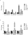
E. cloacae T6SS-1 and T6SS-2 are differentially expressed in TSB and DMEM. Fold change expression (RT-qPCR) of T6SS-1 (A) and T6SS-2 genes (B), compared with LB medium. E. cloacae strains were grown in different culture media: lysogenic broth (LB), trypticase soy broth (TSB), Dulbecco’s Modified Eagle’s Medium (DMEM) with high glucose (4.5 g/l), pleuropneumoniae-like organisms (PPLO) broth, and colonization factor antigen (CFA) broth. Bacterial cultures were grown at 37°C for 6 h. 16S rRNA was used as a reference gene for normalization. Data represent the mean of three independent experiments performed in triplicate. Statistically significant with respect to the WT bacteria grown in LB medium; ns: not significant; ***p < 0.001.
FIGURE 3
FIGURE 3
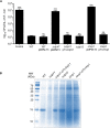
E. cloacae T6SS-1 is required for the bacterial competition. (A) Comparison of the survival of E. coli DH5α against wild-type E. cloacae and ΔclpV1, ΔclpV2, and Δhcp1 mutants. Survival rates are expressed in CFU/ml. Control: E. coli DH5α in LB with no E. cloacae strains. (B) Hcp1 protein was detected by 16% SDS-PAGE and Coomasie Blue staining, from concentrated supernatants of E. cloacae strains grown at 6 h in TSB medium. Statistically significant with respect to the wild-type strain; ns: not significant; ***p < 0.001.
FIGURE 4
FIGURE 4
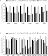
E. cloacae bacterial competition against different enteric bacteria. Comparison of the survival of the different E. coli pathotypes (A) and other Gram-negative pathogens (B) against wild-type E. cloacae (−/+pMPMT-3), Δhcp1 pMPM-T3, and Δhcp1 pT3-Hcp1. EPEC: enteropathogenic E. coli. EHEC: enterohemorragic E. coli. ETEC: enterotoxigenic E. coli. EAEC: enteroaggregative E. coli. APEC: avian-pathogenic E. coli. UPEC: uropathogenic E. coli. KOX: K. oxytoca. KPN: K. pneumoniae. STY: S. Typhi. STM: S. Typhimurium. ECL11E, ECL18E, ECL31E, and ECL43E: clinical isolates of E. cloacae. Survival levels are expressed in CFU/ml. Control: prey bacteria in LB with no E. cloacae strains. Statistically significant with respect to the wild-type strain; ns: not significant; *p < 0.05; **p < 0.01; ***p < 0.001.
FIGURE 5
FIGURE 5
Hemolysis assay describing T6SS-1-mediated hemolytic activity of E. cloacae. Hemolysis of wild-type E. cloacae (−/+pMPM-T3), ΔclpV1 pMPM-T3, ΔclpV1 pT3-ClpV1, ΔclpV2, Δhcp1 pMPM-T3, and Δhcp1 pT3-Hcp backgrounds was quantified by measuring absorbance at 450 nm and expressed in percentage with respect to the lysis obtained with wild-type E. cloacae strain. Statistically significant with respect to the wild-type strain; ns: not significant; *p < 0.05.
FIGURE 6
FIGURE 6
Role of E. cloacae T6SSs on the biofilm formation and cell adherence. (A) Quantification of biofilm formation by measuring crystal violet uptake. Wild-type E. cloacae and T6SS mutants were grown for 24 h in DMEM. (B) Adherence of wild-type E. cloacae ΔclpV1, ΔclpV2, ΔclpV2 pT3-ClpV2, and ΔclpV1 ΔclpV2 backgrounds, after 2 h of infection of HeLa cell monolayers. Statistically significant differences between wild-type E. cloacae and their respective T6SS isogenic mutants; ns: not significant; ***p < 0.001.
FIGURE 7
FIGURE 7
Both T6SSs of E. cloacae are required for the gut colonization. BALB/c mice were infected by intragastric inoculation with 109 CFU/ml with the WT strain and their respective isogenic mutants in the ATPases of both T6SSs. Bacterial colonization was assessed after 3 and 6 days post-infection. Statistically significant differences between wild-type E. cloacae and their respective T6SS isogenic mutants; *p < 0.05; ***p < 0.001.

Similar articles

Cited by

References

    1. Ares M. A., Abundes-Gallegos J., Rodriguez-Valverde D., Panunzi L. G., Jimenez-Galicia C., Jarillo-Quijada M. D., et al. (2019). The Coli Surface Antigen CS3 of Enterotoxigenic Escherichia coli is differentially regulated by H-NS, CRP, and CpxRA Global Regulators. Front. Microbiol. 10:1685. 10.3389/fmicb.2019.01685 - DOI - PMC - PubMed
    1. Ares M. A., Fernandez-Vazquez J. L., Rosales-Reyes R., Jarillo-Quijada M. D., Von Bargen K., Torres J., et al. (2016). H-NS Nucleoid Protein controls virulence features of Klebsiella pneumoniae by regulating the expression of type 3 Pili and the capsule polysaccharide. Front. Cell. Infect. Microbiol. 6:13. 10.3389/fcimb.2016.00013 - DOI - PMC - PubMed
    1. Aschtgen M. S., Bernard C. S., De Bentzmann S., Lloubes R., Cascales E. (2008). SciN is an outer membrane lipoprotein required for type VI secretion in enteroaggregative Escherichia coli. J. Bacteriol. 190 7523–7531. 10.1128/jb.00945-08 - DOI - PMC - PubMed
    1. Barnes A. I., Ortiz C., Paraje M. G., Balanzino L. E., Albesa I. (1997). Purification and characterization of a cytotoxin from Enterobacter cloacae. Can. J. Microbiol. 43 729–733. 10.1139/m97-105 - DOI - PubMed
    1. Basler M., Pilhofer M., Henderson G. P., Jensen G. J., Mekalanos J. J. (2012). Type VI secretion requires a dynamic contractile phage tail-like structure. Nature 483 182–186. 10.1038/nature10846 - DOI - PMC - PubMed

LinkOut - more resources