. 2020 Sep 18:11:573260.
doi: 10.3389/fmicb.2020.573260. eCollection 2020.

Isolation and Characterization of Shewanella Phage Thanatos Infecting and Lysing Shewanella oneidensis and Promoting Nascent Biofilm Formation

Affiliations

Isolation and Characterization of Shewanella Phage Thanatos Infecting and Lysing Shewanella oneidensis and Promoting Nascent Biofilm Formation

Maximilian Kreienbaum et al. Front Microbiol. .

Abstract

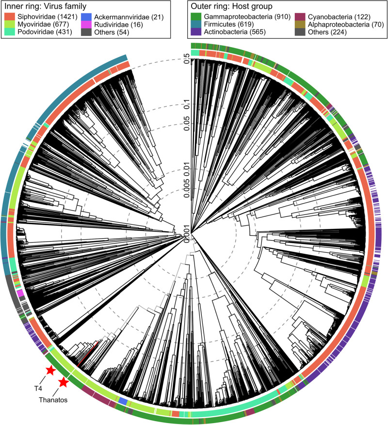
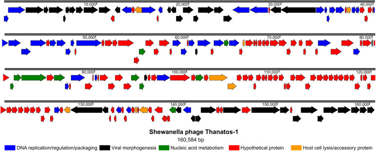
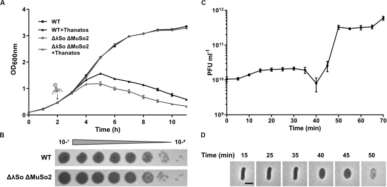
Species of the genus Shewanella are widespread in nature in various habitats, however, little is known about phages affecting Shewanella sp. Here, we report the isolation of phages from diverse freshwater environments that infect and lyse strains of Shewanella oneidensis and other Shewanella sp. Sequence analysis and microscopic imaging strongly indicate that these phages form a so far unclassified genus, now named Shewanella phage Thanatos, which can be positioned within the subfamily of Tevenvirinae (Duplodnaviria; Heunggongvirae; Uroviricota; Caudoviricetes; Caudovirales; Myoviridae; Tevenvirinae). We characterized one member of this group in more detail using S. oneidensis MR-1 as a host. Shewanella phage Thanatos-1 possesses a prolate icosahedral capsule of about 110 nm in height and 70 nm in width and a tail of about 95 nm in length. The dsDNA genome exhibits a GC content of about 34.5%, has a size of 160.6 kbp and encodes about 206 proteins (92 with an annotated putative function) and two tRNAs. Out of those 206, MS analyses identified about 155 phage proteins in PEG-precipitated samples of infected cells. Phage attachment likely requires the outer lipopolysaccharide of S. oneidensis, narrowing the phage's host range. Under the applied conditions, about 20 novel phage particles per cell were produced after a latent period of approximately 40 min, which are stable at a pH range from 4 to 12 and resist temperatures up to 55°C for at least 24 h. Addition of Thanatos to S. oneidensis results in partial dissolution of established biofilms, however, early exposure of planktonic cells to Thanatos significantly enhances biofilm formation. Taken together, we identified a novel genus of Myophages affecting S. oneidensis communities in different ways.

Keywords: LPS; Shewanella; adhesion; biofilm; lysis; phage.


Figures

FIGURE 1
FIGURE 1
Plaque formation and morphology of Shewanella phage Thanatos. (A) Plaques formed by phage Thanatos-1 on agar overlay plates using S. oneidensis ΔLambdaSo ΔMuSo2 as host strain. (B) Cryo electron micrograph of phage Thanatos-1 visualized in a lysed culture of S. oneidensis ΔLambdaSo ΔMuSo2. All isolates showed a highly similar plaque morphology and appearance.
FIGURE 2
FIGURE 2
Viral proteomic tree. Phylogenetic tree created using ViPTree (Nishimura et al., 2017) (available at: https://www.genome.jp/viptree/). The proteomic tree is generated based on genome-wide similarities as determined by tBLASTx. Thanatos phages and all genome sequences of phages from the order Caudovirales from Virus-Host DB (RefSeq release 93) were included in the analysis (ref). Outer and inner ring are colored according to host group and virus family, respectively. For reference, Thanatos phages and Escherichia virus T4 are highlighted by red stars.
FIGURE 3
FIGURE 3
Genome organization of Shewanella phage Thanatos. The image displays the predicted open reading frames within the phage genome indicating their transcriptional direction. The deduced gene products are color-coded as indicated below according to their predicted functional categories. The ORF plot has been generated using SnapGene software (from Insightful Science; available at snapgene.com). For the corresponding annotations see Supplementary Table S3.
FIGURE 4
FIGURE 4
Infection dynamics, latent phase and burst size of Thanatos. (A) Lysis assay during planktonic growth of S. oneidensis WT and ΔLambdaSo ΔMuSo2 infected with Thanatos compared to uninfected cultures. Phages were added 2 h post inoculation (represented by the arrow) at an MOI of 0.1. The experiment was performed in three biological replicates with technical duplicates each. Error bars represent the SEM. (B) Phage spot assay of Thanatos on S. oneidensis WT and ΔLambdaSo ΔMuSo2. The 10– 1 dilution equals a phage concentration of ∼109 PFU ml– 1. (C) One step growth experiment. S. oneidensis ΔLambdaSo ΔMuSo2 was chosen as host strain. The burst size was calculated by dividing the average phage titer at the plateau phase by the average phage titer along the latent phase. Error bars represent the SEM. (D) Time lapse analysis of a S. oneidensis ΔLambdaSo ΔMuSo2 cell infected with Thanatos. The scale bar equals 2 μm.
FIGURE 5
FIGURE 5
Temperature and pH sensitivity of Thanatos. (A) Thermal stability test (24 h). (B) pH stability test (24 h). The undiluted phage preparation contained ∼109 PFU ml– 1. Shewanella oneidensis ΔLambdaSo ΔMuSo2 served as host strain. All images are representatives from three biological replicates.
FIGURE 6
FIGURE 6
Deletion of waaC protects Shewanella oneidensis from Thanatos infection by preventing phage adsorption. (A) Spot Assay of Thanatos on Shewanella oneidensis WT, ΔLambdaSo ΔMuSo2, waaC deletion and waaC complementation strains. The 10– 2 dilution equals a phage concentration of ∼108 PFU ml– 1. (B) Phage adsorption assay. The cells were grown to early exponential phase (OD600 0.15), before 2 × 10⁶ PFU ml– 1 were added to the culture. Samples were taken at the indicated time points and the number of PFU was determined.
FIGURE 7
FIGURE 7
Thanatos differently influences developing and mature biofilms depending on the presence of prophages. Shown is the biofilm formation under static conditions of infected Shewanella oneidensis WT and Shewanella oneidensis ΔLambdaSo ΔMuSo2 cultures compared to uninfected cultures. Values of panels (A–D) represent the percentage of biofilm biomass of treated cultures in comparison to untreated cultures. Biofilm biomass of the untreated cultures was set to 100% (indicated by the blue line). (A,C) Short term biofilm assay. Cultures were inoculated in 96 well microtiter plates at 0 h. The time points indicate the addition of phages to the developing biofilm. Absorption of crystal violet was measured after 24 h. (B,D) Long-term biofilm assay. After inoculation of the cultures, the microtiter plate was incubated without the addition of phages. After 24 h, infectious phages were added to the mature biofilms. Absorption of crystal violet was measured 2, 6, 9 and 24 h after the addition of phages. *p < 0.05; **p < 0.01; ***p < 0.0001. (E) Microscopic analysis of biofilms. 24 h-old biofilms were counterstained with SYTO9 (cyan) and propidium iodide (red). Scale bars = 50 μm. (F) 96-well microtiter plate showing the macroscopic biofilm morphology. A static biofilm assay was performed and the biofilms were documented after removal of crystal violet.

Similar articles

Cited by

References

    1. Ackermann H. W. (2009). Phage classification and characterization. Methods Mol. Biol. 501 127–140. 10.1007/978-1-60327-164-6_13 - DOI - PubMed
    1. Adams M. (1959). Bacteriophages. New York, NY: Interscience Publishers.
    1. Argov T., Azulay G., Pasechnek A., Stadnyuk O., Ran-Sapir S., Borovok I., et al. (2017). Temperate bacteriophages as regulators of host behavior. Curr. Opin. Microbiol. 38 81–87. 10.1016/j.mib.2017.05.002 - DOI - PubMed
    1. Beblawy S., Bursac T., Paquete C., Louro R., Clarke T. A., Gescher J. (2018). Extracellular reduction of solid electron acceptors by Shewanella oneidensis. Mol. Microbiol. 109 571–583. 10.1111/mmi.14067 - DOI - PubMed
    1. Binnenkade L., Teichmann L., Thormann K. M. (2014). Iron triggers lambdaSo prophage induction and release of extracellular DNA in Shewanella oneidensis MR-1 biofilms. Appl. Environ. Microbiol. 80 5304–5316. 10.1128/AEM.01480-14 - DOI - PMC - PubMed