. 2020 Sep 23:11:551413.
doi: 10.3389/fimmu.2020.551413. eCollection 2020.

Adenovirus 7 Induces Interlukin-6 Expression in Human Airway Epithelial Cells via p38/NF-κB Signaling Pathway

Affiliations

Adenovirus 7 Induces Interlukin-6 Expression in Human Airway Epithelial Cells via p38/NF-κB Signaling Pathway

Lifeng Qi et al. Front Immunol. .

Abstract

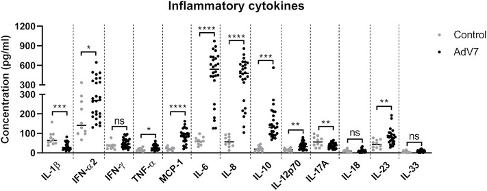
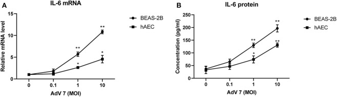
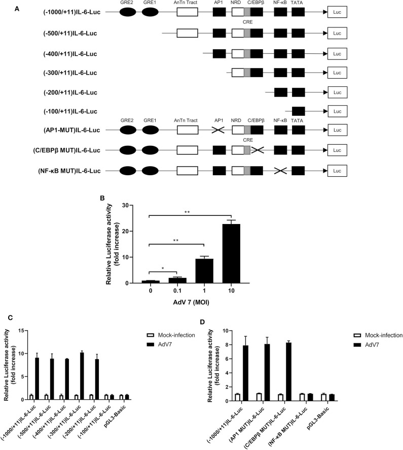
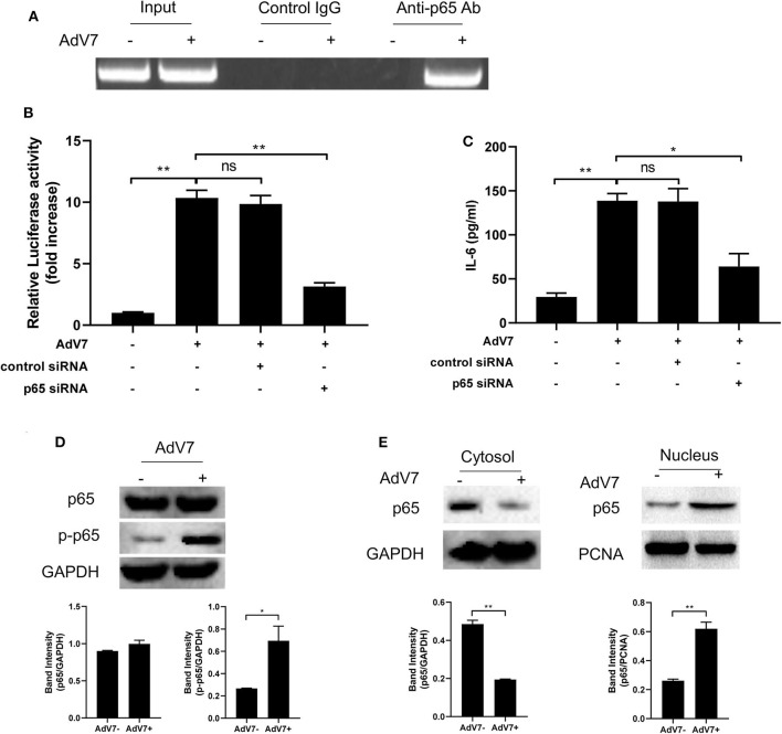
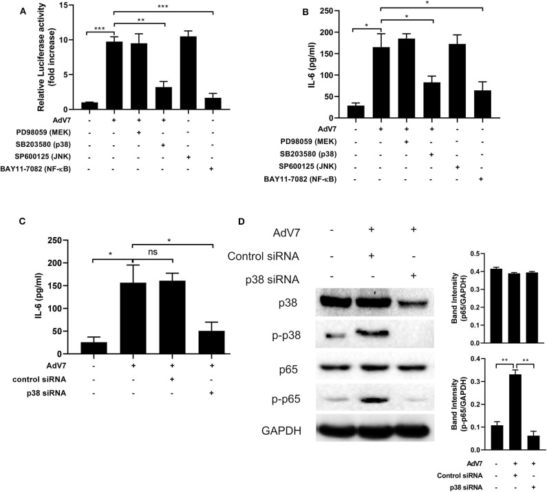
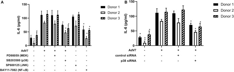
Human Adenovirus (AdV) infection is very common and usually has a significant impact on children. AdV-induced inflammation is believed to be one of the main causes of severe symptoms. However, an inflammatory response profile in the airway in AdV-infected children is still lacking, and the mechanism underlying AdV-induced inflammation in the airway is also poorly understood. In the current study, we determined the expression of a panel of inflammation cytokines in the airway samples from AdV 7 infected children and further investigated the molecular mechanism underlying AdV 7-induced cytokine expression. Our results showed that eight out of 13 tested inflammatory cytokines were significantly increased in nasal washes of AdV 7-infected children comparing to healthy control, with IL-6 showing the highest enhancement. AdV 7 infection of bronchial epithelial cell line and primary airway epithelial cells confirmed that AdV 7 increased IL-6 mRNA and protein expression in an infection dose-dependent manner. Promoter analysis revealed that AdV 7 infection transactivated IL-6 promoter and a NF-κB binding site in IL-6 promoter was involved in the transactivation. Further analysis showed that upon AdV 7 infection, NF-κB p65 was phosphorylated and translocated into nucleus and bound onto IL-6 promoter. Signaling pathway analysis revealed that p38/NF-κB pathway was involved in AdV 7 infection induced IL-6 elevation. Taken together, our study shows that AdV 7 infection triggers the expression of a range of inflammatory cytokines including IL-6 in the airway of infected children, and AdV 7 enhances IL-6 expression by transactivating IL-6 promoter via p38/NF-κB signaling pathway. Findings of our current study have provided more information toward a better understanding of AdV-induced airway inflammation, which might also benefit the development of intervention strategies.

Keywords: Adenovirus 7; Interlukin-6; airway epithelial cell; inflammation; p38/NF-κB signaling pathway.


Figures

Figure 1
Figure 1
AdV 7 infection triggers the production of inflammatory cytokines in the airway in children. Nasal samples from AdV 7 infected and healthy children were harvested and inflammatory cytokines IL-1β, IFN-α2, IFN-γ, TNF-α, MCP-1, IL-6, IL-8, IL-10, IL-12p70, IL-17A, IL-18, IL-23, and IL-33 were measured by BioLegend's LEGENDplex bead-based immunoassay. ns, statistically not significant; *p < 0.05; **p < 0.01; ***p < 0.001; ****p < 0.0001.
Figure 2
Figure 2
AdV 7 infection induces IL-6 mRNA and protein expression in airway epithelial cells. Human normal bronchial epithelial cell line BEAS-2B and primary human airway epithelial cells (hAEC) were either mock-infected or infected with ascendant doses of AdV 7 for 24 h and then (A) IL-6 mRNA and (B) protein expression were measured by RT-PCR and ELISA, respectively. Data shown are mean ± SD of three independent experiments with each condition performed in duplicate. *p < 0.05; **p < 0.01.
Figure 3
Figure 3
A NF-κB binding site in the IL-6 promoter is involved in AdV 7-induced IL-6 transactivation. (A) Schematic illustration of all the IL-6 promoter-controlled luciferase reporter gene plasmids. (B) BEAS-2B cells were first transfected with (-1,000/+11)IL-6-Luc, and then either mock-infected or infected with ascendant doses of AdV 7 for 24 h. Afterwards, cells were lysed and luciferase activity was measured. Data shown are mean ± SD of three independent experiments with each condition performed in duplicate. *p < 0.05; **p < 0.01. (C,D) BEAS-2B cells were first infected with either (C) serially truncated IL-6 promoter plasmids or (D) site-directed mutated IL-6 promoter plasmids, and then were either mock-infected or infected with 1 MOI of AdV 7. Twenty-four hours later, cells were lysed, and luciferase activity was measure. Data shown are mean ± SD of three independent experiments with each condition performed in duplicate.
Figure 4
Figure 4
AdV 7 infection induces NF-κB binding onto IL-6 promoter. (A) BEAS-2B cells were either mock-infected or infected with 1 MOI AdV 7 for 24 h, and then ChIP assay was performed with either control IgG or anti-p65 antibody. The amplification of IL-6 promoter sequence from ChIP-pulldown was performed with PCR. One representative result is shown. (B) BEAS-2B cells were sequentially transfected with control siRNA or p65 siRNA and (-1,000/+11)IL-6-Luc, and then were mock-infected or infected with 1 MOI AdV 7. Twenty-four hours later, cells were lysed, and luciferase activity was measured. Data shown are mean ± SD of three independent experiments with each condition performed in duplicate. Ns, not statistically significant; **p < 0.01. (C) BEAS-2B cells were first transfected with either control siRNA or p65 siRNA, and then were mock-infected or infected with 1 MOI AdV 7. Twenty-four hours later, IL-6 concentration in the cell culture supernatant was quantified by ELISA. Data shown are mean ± SD of three independent experiments with each condition performed in duplicate. Ns, not statistically significant; *p < 0.05; **p < 0.01. (D,E) BEAS-2B cells were either mock-infected or infected with 1 MOI AdV 7, and then (D) cells were lysed, or (E) cell cytoplasmic and nuclear fractions were isolated, and then (D,E) the expression of p65 and p-p65 was determined by Western blot. One out of three results is sown.
Figure 5
Figure 5
AdV 7 infection induces IL-6 expression via p38/NF-κB signaling pathway. (A,B) BEAS-2B cells were first (A) transfected with (-1,000/+11)IL-6-Luc and then (A,B) mock-infected or infected with 1 MOI AdV 7 and treated with different signaling pathway inhibitors. Twenty-four hours later, (A) cells were lysed and luciferase activity was measured, or (B) IL-6 concentration in the supernatant was measured by ELISA. Data shown are mean ± SD of three independent experiments with each condition performed in duplicate. *p < 0.05; **p < 0.01; ***p < 0.001. (C,D) BEAS-2B cells were first transfected with control siRNA or p38 siRNA, and then were mock-infected or infected with 1 MOI AdV 7. Twenty-four hours later, (C) IL-6 concentration in the supernatant was measured by ELISA, or (D) cells were lysed and the expression of p38, p-p38, p65, and p-p65 was determined by Western blot. (C) Data shown are mean ± SD of three independent experiments with each condition performed in duplicate. Ns, not statistically significant; *p < 0.05. (D) One representative result out of three is shown.
Figure 6
Figure 6
Blocking p38/NF-κB signaling pathway suppresses AdV 7-induced IL-6 expression. (A,B) primary hAEC cells were first (A) treated with signaling pathway inhibitors or (B) transfected with control siRNA or p38 siRNA, and then (A,B) infected with 1 MOI AdV 7. Twenty-four hours later, IL-6 concentration was quantified by ELISA. Data shown are mean ± SD of three independent experiments with each condition performed in duplicate. All statistical comparisons were made against cells with AdV 7 infection only. Ns, not statistically significant; *p < 0.05; **p < 0.01.

Similar articles

Cited by

References

    1. Xie L, Zhang B, Xiao N, Zhang F, Zhao X, Liu Q, et al. . Epidemiology of human adenovirus infection in children hospitalized with lower respiratory tract infections in Hunan, China. J Med Virol. (2019) 91:392–400. 10.1002/jmv.25333 - DOI - PMC - PubMed
    1. Moro MR, Bonville CA, Suryadevara M, Cummings E, Faddoul D, Kobayaa H, et al. . Clinical features, adenovirus types, and local production of inflammatory mediators in adenovirus infections. Pediatric Infect Dis J. (2009) 28:376–80. 10.1097/INF.0b013e31819075a6 - DOI - PubMed
    1. Chen R-F, Lee C-Y. Adenoviruses types, cell receptors and local innate cytokines in adenovirus infection. Int Rev Immunol. (2014) 33:45–53. 10.3109/08830185.2013.823420 - DOI - PubMed
    1. Becroft D. Histopathology of fatal adenovirus infection of the respiratory tract in young children. J Clin Pathol. (1967) 20:561–9. 10.1136/jcp.20.4.561 - DOI - PMC - PubMed
    1. Chuang Y, Chiu C-H, Wong K-S, Huang J-G, Huang YC, Chang LY, et al. . Severe adenovirus infection in children. J Microbiol Immunol Infect. (2003) 36:37–40. - PubMed

Publication types

MeSH terms

LinkOut - more resources