. 2020 Oct 15:17:86.
doi: 10.1186/s12986-020-00508-1. eCollection 2020.

High-protein and low-calorie diets improved the anti-aging Klotho protein in the rats' brain: the toxic role of high-fat diet

Affiliations

High-protein and low-calorie diets improved the anti-aging Klotho protein in the rats' brain: the toxic role of high-fat diet

Anahid Shafie et al. Nutr Metab (Lond). .

Abstract

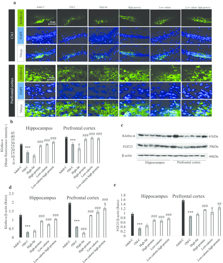
Background: In the current study, our specific aim was to characterize the Klotho protein and expression levels in the hippocampus and prefrontal cortex of old rats treated with different diets (high-fat, high-protein, low-calorie, high-protein and low-calorie).

Methods: Rats were treated with high-fat, high-protein, low-calorie, low-calorie high-protein diets for 10 weeks and then behavioral and molecular assessments were evaluated.

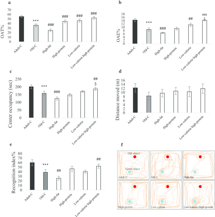
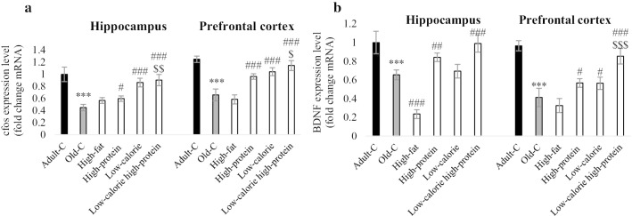
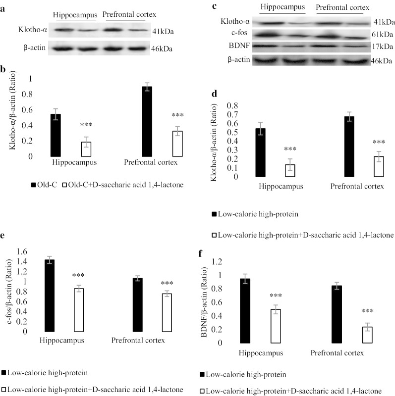
Results: Statistical analysis showed the percentage of open arm time was increased in the high-protein, low-calorie and low-calorie high-protein groups compared with old control (old-C) rats. The percentage of open arm entries was increased in the low-calorie and low-calorie high-protein group compared with old-C rats. The body weight and serum triglyceride were decreased in the low-calorie and low-calorie high-protein groups in comparison to control old rats. Low-calorie and low-calorie high-protein treatments statistically enhanced caspase-3 level compared with old-C rats in the hippocampus and prefrontal cortex. Treatment of old rats with high-protein, low-calorie and low-calorie high-protein could increase Klotho-α level compared with control old rats. The levels of Klotho-α, c-fos and brain-derived neurotrophic factors were decreased in the low-calorie high-protein group in Klotho inhibitor's presence compared with the low-calorie high-protein group.

Conclusion: According to our findings, Klotho-α level was reduced in old rats. Low-calorie, high-protein and particularly low-calorie high-protein diets increased this protein level and consequently increased neuronal plasticity and improved memory function.

Keywords: FGF23; High-protein diet; Klotho; Low-calorie diet; c-fos.


Conflict of interest statement

Competing interestsThe authors declare they have no conflict of interests.

Figures

Fig. 1
Fig. 1
Behavioral assessments. Rats treated with high-fat, high-protein, low-calorie, low-calorie high-protein diets for 10 weeks. Control adult and old rats treated with rodent standard pellet. After 10 weeks, behavioral tests were performed (n = 7). Anxiety-like behavior was assessed by elevated plus maze test (EPM) for 5 min. Percentage of open arm time (a) and percentage of open arm entires (b) in the EPM were shown. Center occupancy (d) and locomotion (c) were assessed by open field test for 5 min (n = 7). Percentage of recognition index (e) was measured by novel object recognition test (n = 7). A represented tracks showing 2 min path in the NOR (f). Data were presented as Mean ± S.E.M. ***P < 0.001 ver. Adult-C, ##P < 0.01, ###P < 0.001 ver. Old-C. $P < 0.05 ver. High-protein. Adult-C adult control rats, Old-C old control rats
Fig. 2
Fig. 2
Molecular assessment of Klotho-α and FGF23 in the hippocampus and prefrontal cortex of experimental groups. Rats treated with high-fat, high-protein, low-calorie, low-calorie high-protein diets for 10 weeks. Control adult and old rats treated with rodent standard pellet. After behavioral tests, rats were euthanized and brains of three of them perfused for histological evaluation in each group (n = 3) and brains of other three rats in each groups were collected for Western blotting technique (n = 4). Immunoflorence assay a showed the Klotho-α distribution in the hippocampus and prefrontal cortex. Mean of fluorescence intensity showed Klotho-α positive cell in the CA1 and prefrontal cortex (b). A represented blot showed Klotho-α and FGF23 protein level (c) and the density of Klotho-α bands (d) and FGF23 band (e) were measured in the hippocampus and prefrontal cortex (n = 4 and technical repeat for each n is 3). Data were presented as Mean ± S.D. ***P < 0.001 ver. Adult-C, #P < 0.05, ##P < 0.01, ###P < 0.001 ver. Old-C. $P < 0.05 ver. High-protein. Adult-C adult control rats, Old-C old control rats
Fig. 3
Fig. 3
Gene expression of c-fos (a) and BDNF (b) in the hippocampus and prefrontal cortex (n = 4). Rats treated with high-fat, high-protein, low-calorie, low-calorie high-protein diets for 10 weeks. Control adult and old rats treated with rodent standard pellet. After behavioral tests, rats were euthanized and brains of three rats in each groups were collected for RT-PCR (n = 4 and technical repeat for each n is 3). Data were presented as Mean ± S.D. ***P < 0.001 ver. Adult-C, #P < 0.05, ##P < 0.01, ###P < 0.001 ver. Old-C. $P < 0.05, $$P < 0.01, $$$P < 0.001 ver. High-protein. Adult-C adult control rats, Old-C old control rats
Fig. 4
Fig. 4
Molecular assessment of Klotho-α, c-fos and BDNF in the hippocampus and prefrontal cortex of experimental groups (n = 4). A represented blot showed Klotho-α in the old rats with standard diet for 10 weeks and old rats with Klotho-α inhibitor (d-saccharic acid 1,4-lactone, orally administrated 2 time/week for 10 weeks) (a) and the density of Klotho-α band (b) was measured. A represented blot showed Klotho-α, c-fos and BDNF in the low-calorie high-protein group (treated for 10 weeks) with and without Klotho-α inhibitor (c) and the density of Klotho-α (d), c-fos (e) and BDNF (f) bands were measured in the hippocampus and prefrontal cortex. Data were presented as Mean ± S.D. ***P < 0.001 ver. low-calorie high-protein group. Adult-C adult control rats, Old-C old control rats

Similar articles

Cited by

References

    1. Mehta HH, Xiao JL, Ramirez R, Miller B, Kim SJ, et al. Metabolomic profile of diet-induced obesity mice in response to humanin and small humanin-like peptide 2 treatment. Metabolomics. 2019;15:88. doi: 10.1007/s11306-019-1549-7. - DOI - PMC - PubMed
    1. Paoli A, Bianco A, Damiani E, Bosco G. Ketogenic diet in neuromuscular and neurodegenerative diseases. Biomed Res Int. 2014;2014:474296. doi: 10.1155/2014/474296. - DOI - PMC - PubMed
    1. Mero AA, Huovinen H, Matintupa O, Hulmi JJ, Puurtinen R, et al. Moderate energy restriction with high protein diet results in healthier outcome in women. J Int Soc Sports Nutr. 2010;7:4. doi: 10.1186/1550-2783-7-4. - DOI - PMC - PubMed
    1. Kurosu H, Yamamoto M, Clark JD, Pastor JV, Nandi A, et al. Suppression of aging in mice by the hormone Klotho. Science. 2005;309:1829–1833. doi: 10.1126/science.1112766. - DOI - PMC - PubMed
    1. Kuroo M, Matsumura Y, Aizawa H, Kawaguchi H, Suga T, et al. Mutation of the mouse klotho gene leads to a syndrome resembling ageing. Nature. 1997;390:45–51. doi: 10.1038/36285. - DOI - PubMed