. 2020 Oct 7;9(10):e1189.
doi: 10.1002/cti2.1189. eCollection 2020.

Kinetics and isotype assessment of antibodies targeting the spike protein receptor-binding domain of severe acute respiratory syndrome-coronavirus-2 in COVID-19 patients as a function of age, biological sex and disease severity

Affiliations

Kinetics and isotype assessment of antibodies targeting the spike protein receptor-binding domain of severe acute respiratory syndrome-coronavirus-2 in COVID-19 patients as a function of age, biological sex and disease severity

Nancy R Graham et al. Clin Transl Immunology. .

Abstract

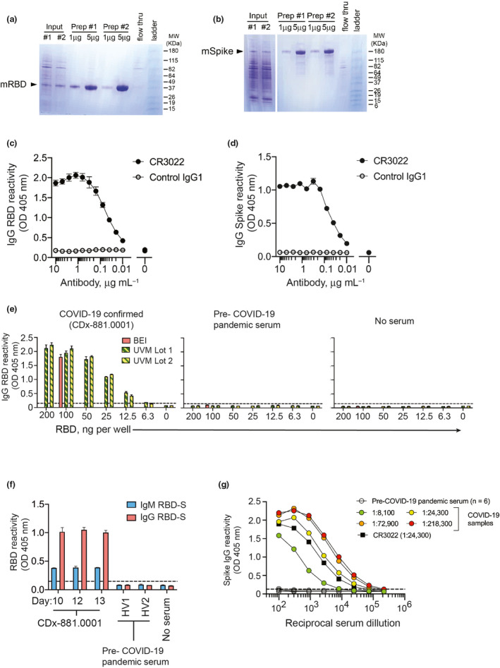
Objectives: There is an incomplete understanding of the host humoral immune response to severe acute respiratory syndrome (SARS)-coronavirus (CoV)-2, which underlies COVID-19, during acute infection. Host factors such as age and sex as well as the kinetics and functionality of antibody responses are important factors to consider as vaccine development proceeds. The receptor-binding domain of the CoV spike (RBD-S) protein mediates host cell binding and infection and is a major target for vaccine design to elicit neutralising antibodies.

Methods: We assessed serum anti-SARS-CoV-2 RBD-S IgG, IgM and IgA antibodies by a two-step ELISA and neutralising antibodies in a cross-sectional study of hospitalised COVID-19 patients of varying disease severities. Anti-RBD-S IgG levels were also determined in asymptomatic seropositives.

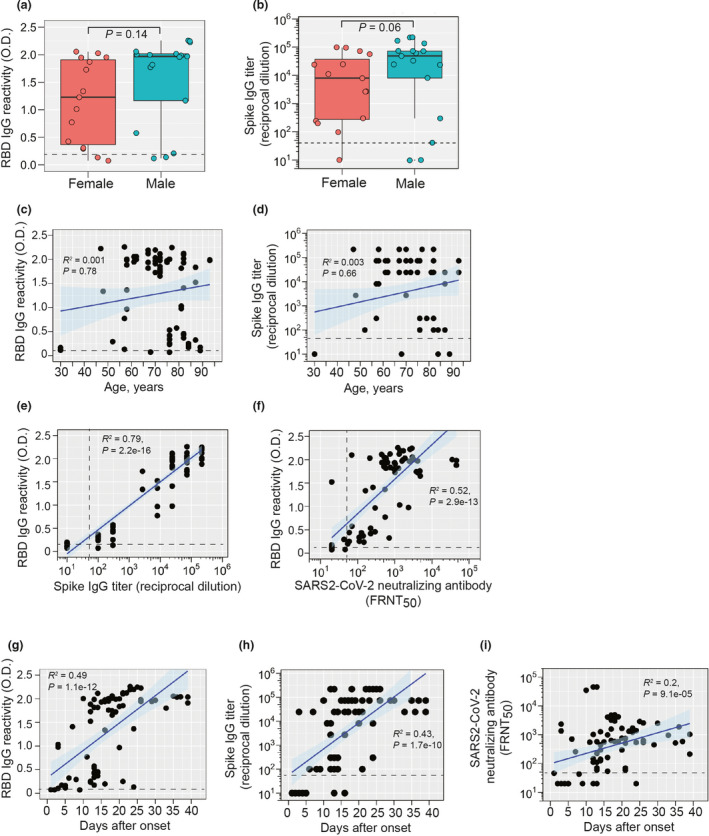
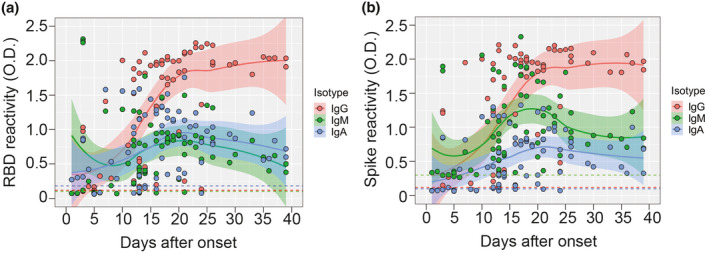
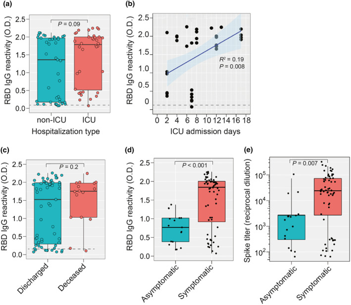
Results: We found equivalent levels of anti-RBD-S antibodies in male and female patients and no age-related deficiencies even out to 93 years of age. The anti-RBD-S response was evident as little as 6 days after onset of symptoms and for at least 5 weeks after symptom onset. Anti-RBD-S IgG, IgM and IgA responses were simultaneously induced within 10 days after onset, with anti-RBD-S IgG sustained over a 5-week period. Anti-RBD-S antibodies strongly correlated with neutralising activity. Lastly, anti-RBD-S IgG responses were higher in symptomatic COVID-19 patients during acute infection compared with asymptomatic seropositive donors.

Conclusion: Our results suggest that anti-RBD-S IgG reflect functional immune responses to SARS-CoV-2, but do not completely explain age- and sex-related disparities in COVID-19 fatalities.

Keywords: COVID‐19; SARS‐CoV‐2; humoral immune response; isotypes; neutralising antibody; spike protein.


Conflict of interest statement

The authors declare no conflict of interest.

Figures

Figure 1
Figure 1
Validation of SARS‐CoV‐2 RBD‐S and spike antigens in COVID‐19 samples. Reducing SDS‐PAGE analysis of (a) RBD‐S and (b) trimeric spike purified from transiently transfected mammalian HEK293 cells. (c) Binding of CR3022 IgG1 mAb to SARS‐CoV‐2 RBD‐S and (d) trimerised spike. The anti‐dengue virus 1M7 IgG1 mAb 42 was used as a control. (e) Detection of serum IgG from a COVID‐19 patient (left), but not from pre‐2020 serum (centre) or no serum control (right). (f) Detection of IgM and IgG to RBD‐S in serial time the course serum samples from a COVID‐19 patient and not in pre‐COVID‐19 pandemic healthy volunteer sera (HV1,HV2). All sera diluted 1:50, and for the COVID‐19 patient (CD×‐881.0001), day after symptoms onset is shown on the x‐axis. (g) Anti‐spike IgG reactivity by ELISA for pre‐COVID‐19 pandemic sera (n = 6), the anti‐SARS‐CoV‐1/2 mAb CR3022, and COVID‐19 samples of varying titres (indicated in parentheses).
Figure 2
Figure 2
IgG responses to SARS‐CoV‐2 RBD‐S and spike. (a) Comparison of RBD‐S IgG reactivity (OD 405 nm) or (b) anti‐spike IgG endpoint titres levels in male (n = 17) or female (n = 15) patients. Some patients had multiple samples that were averaged for this analysis. Boxplots show the 25–75th percentiles, with median as horizontal line and whiskers as 95% confidence level with subjects as symbols and groups were analysed by Mann–Whitney U‐tests. All patient samples were plotted individually. (c) Anti‐RBD‐S IgG and (d) anti‐spike IgG are expressed as a function of age with all individual samples from a patient plotted (n = 77 total). (e) Anti‐RBD‐S IgG reactivity is plotted against anti‐spike IgG endpoint titres. (f) SARS‐CoV‐2 microneutralisation titres [50% focus reduction neutralisation titres (FRNT50)] are plotted against RBD‐S IgG reactivity (n = 77). (g) RBD‐S IgG reactivity, (h) anti‐spike IgG endpoint titres, and (i) SARS‐CoV‐2 neutralising antibody titres are plotted against days after symptoms onset. (Cut‐off values for each assay are shown by dashed lines. Spearman's Rho coefficient (R 2), 95% confidence interval (shading), and P‐values are shown for panels c–i).
Figure 3
Figure 3
Antibody isotype usage during the response to SARS‐CoV‐2 RBD‐S and spike. (a) RBD‐S IgM, IgG and IgA in serum (diluted 1:50) were determined by ELISA and plotted against days post‐onset of symptoms (n = 77 samples). LOESS‐smoothed lines and 95% confidence intervals are shown for each isotype. (b) Spike‐reactive IgM, IgG and IgA in serum (diluted 1:100) were determined by ELISA and plotted against days post‐onset of symptoms. LOESS‐smoothed lines and 95% confidence intervals are shown for each isotype.
Figure 4
Figure 4
SARS‐CoV‐2 RBD‐S IgG responses during hospitalisation. (a) RBD‐S IgG in patients that were hospitalised in the ICU or not. (b) For ICU‐hospitalised patients, all RBD‐S IgG values are presented as a function of ICU admission days. Spearman's Rho coefficient (R 2), 95% confidence interval, and P‐value are shown. (c) RBD‐S IgG in patients that were deceased (n = 16 samples from seven patients) or discharged (n = 61 samples from 25 patients). (d) RBD‐S IgG reactivity and (e) anti‐S IgG titres in symptomatic COVID‐19 patients (n = 77 samples) versus convalescent seropositive COVID‐19‐exposed volunteers (n = 17). Boxplots in a, c–e show the median, 95% confidence level and all individual samples. The Student's t‐test P‐value is shown.

Update of

References

    1. Cui J, Li F, Shi ZL. Origin and evolution of pathogenic coronaviruses. Nat Rev Microbiol 2019; 17: 181–192. - PMC - PubMed
    1. Lu R, Zhao X, Li J et al Genomic characterisation and epidemiology of 2019 novel coronavirus: implications for virus origins and receptor binding. Lancet 2020; 395: 565–574. - PMC - PubMed
    1. Zhou P, Yang XL, Wang XG et al A pneumonia outbreak associated with a new coronavirus of probable bat origin. Nature 2020; 579: 270–273. - PMC - PubMed
    1. Hoffmann M, Kleine‐Weber H, Schroeder S et al SARS‐CoV‐2 cell entry depends on ACE2 and TMPRSS2 and is blocked by a clinically proven protease inhibitor. Cell 2020; 181: 271–280. - PMC - PubMed
    1. Wan Y, Shang J, Graham R, Baric RS, Li F. Receptor recognition by the novel coronavirus from Wuhan: an analysis based on decade‐long structural studies of SARS coronavirus. J Virol 2020; 94: e00127‐20. - PMC - PubMed