Review
. 2020 Oct 13:6:48.
doi: 10.1186/s40942-020-00250-z. eCollection 2020.

The Tie2 signaling pathway in retinal vascular diseases: a novel therapeutic target in the eye

Affiliations
Review

The Tie2 signaling pathway in retinal vascular diseases: a novel therapeutic target in the eye

Quan Dong Nguyen et al. Int J Retina Vitreous. .

Abstract

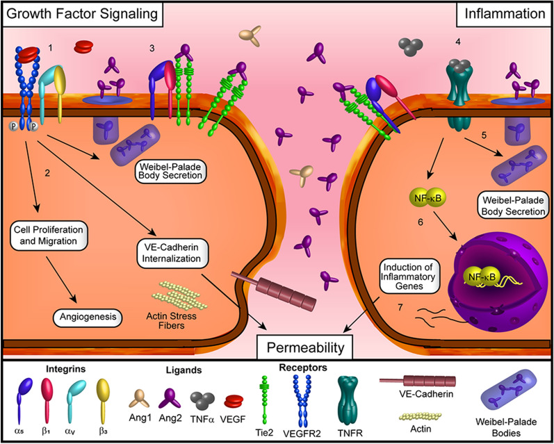
Background: Retinal vascular diseases such as neovascular age-related macular degeneration, diabetic retinopathy and/or diabetic macular edema, and retinal vein occlusion with macular edema-share several key pathophysiologic aspects including neovascularization, vascular permeability, and inflammation. The role of vascular endothelial growth factor (VEGF) in these processes, and the therapeutic benefits of VEGF inhibition, have been well characterized. Anti-VEGF therapy is highly effective for many patients but is not uniformly effective in all patients and imposes a significant treatment burden. More recently, the role of the Tie2 signaling pathway in the pathophysiology of retinal vascular diseases has been investigated, and the Tie2 pathway represents a novel therapeutic target for these conditions.

Areas covered: The index review describes the Tie2 pathway and its complementary role to the VEGF pathway in the angiogenesis cascade and will summarize studies of molecules in development to therapeutically modulate the Tie2 pathway in retinal vascular diseases.

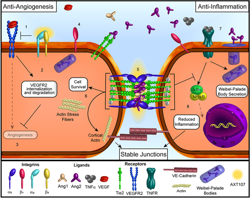
Conclusions: Activation of the Tie2 pathway leads to downstream signaling that promotes vascular health and stability and decreases vascular permeability and inflammation. AXT107 is a collagen IV-derived synthetic peptide with a dual mechanism of action that involves suppression of VEGF signaling and activation of the Tie2 pathway; these actions are accomplished by AXT107 binding to and disrupting different integrin, leading to blockade of the VEGF receptor and rearrangement of cellular Tie2 rendering it susceptible to Ang2 agonism. Other Tie2 agonist compounds are also in development, including faricimab and razuprotafib. Tie2 activation only modestly impacts angiogenesis on its own but significantly potentiates VEGF suppression. Co-regulation of the VEGF and Tie2 signaling pathways has the potential to improve functional and structural outcomes in eyes with retinal vascular diseases.

Keywords: AXT107; Integrin; Tie2; Vascular endothelial grwoth factor; Vascular permeability.


Conflict of interest statement

Competing interestsQDN serves on the Scientific Advisory Board for AbbVie, Bayer, Genentech, Mallinckrodt, Regeneron, and Santen, among others and advises on Drug Safety for AsclepiX; QDN also chaired the Steering Committee for the RIDE/RISE and STOP-Uveitis Study and was on the Steering Committee for other studies sponsored by Genentech and Regeneron. JSH serves on the Scientific Advisory Board for 4DMT, Adverum, Aerie, Aerpio, Akros, Allegro, Apellis, Array, Asclepix, Bayer, Beaver-Visitec, BioMarin, Clearside, Corcept, Daiichi Sankyo, Galecto, Galimedix, Genentech/Roche, Helio, Hemera, Interface, iRenix, Janssen, Kanghong, Kodiak, Notal Vision, Novartis, Ocular Therapeutix, Optos, Orbit Biomedical, Quark, Ra Pharmaceuticals, Regeneron, REGENXBIO, Santen, Scifluor, Shire, Spark Therapeutics, Stealth, Thrombogenics, Tyrogenex. DVD serves on the Scientific Advisory Board for Allergan, AsclepiX, Genentech, Kodiak, Regeneron, and Santen and she has received research support from Genentech and Regeneron. ACM, NBP, HS, and TH are employees of AsclepiX Therapeutics.

Figures

Fig. 1
Fig. 1
VEGF and TNF signaling in retinal vascular disease. Increased expression of angiogenic and inflammatory factors (such as VEGF and TNFα) are associated with retinal vascular diseases. (1) The binding of VEGF to VEGFR2 in complex with αvβ3 integrin stimulates the autophosphorylation of VEGFR2 and the activation of downstream cellular processes. (2) These processes include the induction of migration and proliferation important for angiogenesis and the reorganization of diffuse actin into stress fibers and the internalization of cell–cell adhesion proteins that result in increased permeability. (3) VEGFR2 signaling also stimulates the release of Ang2 from Weibel-Palade bodies, which then competes with Ang1 to bind to Tie2 and reduce the associated signaling pathways related to vascular stability and anti-permeability. (4) With respect to inflammatory signaling, the binding of TNFα to the TNFR also stimulates the release of Ang2 from Weibel-Palade bodies (5) and reduces the activation of Tie2. This not only reduces the vessel stabilizing pathways mentioned above but also blocks the anti-inflammatory activity of the Tie2 receptor as well. (6) The activation of the TNFR pathway by TNFα also stimulates the relocation of NF-κB to the nucleus where it functions as a transcription factor to induce the expression of numerous inflammation-associated genes. (7) Among other functions, the initiation of inflammation stimulates increased permeability within blood vessels
Fig. 2
Fig. 2
Effects of AXT107-treatment in retinal vascular disease. (1) AXT107 binds to αvβ3 integrins and dissociates them from VEGFR2. (2) The disruption of these interactions directly inhibits the autophosphorylation of VEGFR2 in the presence of VEGF and further reduces VEGFR2 responses by increasing the internalization and degradation of the receptor, overall reducing the angiogenesis responses and induction of vessel permeability (3). (4) AXT107 also binds to α5β1 integrin heterodimers, which associate with Tie2, and dissociates the subunits. (5) α5 integrin and Tie2 then relocate to the endothelial cell–cell junctions and form clusters that can be activated following the binding of either Ang1 or Ang2. (6) Active Tie2 clusters stimulate downstream pathways associated with improved endothelial cell survival and the reorganization of intracellular actin from stress fibers into cortical actin that is distributed around the edges of the cell to stabilize the vasculature. (7) Initial activation of TNFR by TNFα does not seem to be affected by AXT107 treatment, resulting in the release of Ang2 from Weibel-Palade bodies. The released Ang2 contributes to the activation of Tie2 clusters formed following AXT107 treatment. (8) Signaling by the phosphorylated Tie2 clusters prevent the degradation of IκBα molecules, retaining NF-κB within the cytoplasm and blocking the induction of inflammation and the associated effects on vessel permeability, thereby contributing to increased vessel stability

Similar articles

Cited by

References

    1. Shweiki D, Itin A, Soffer D, Keshet E. Vascular endothelial growth factor induced by hypoxia may mediate hypoxia-initiated angiogenesis. Nature. 1992;359:843–845. doi: 10.1038/359843a0. - DOI - PubMed
    1. Ferrara N. Vegf and intraocular neovascularization: from discovery to therapy. Transl Vis Sci Technol. 2016;5:10. doi: 10.1167/tvst.5.2.10. - DOI - PMC - PubMed
    1. Aiello LP, Avery RL, Arrigg PG, et al. Vascular endothelial growth factor in ocular fluid of patients with diabetic retinopathy and other retinal disorders. N Engl J Med. 1994;331:1480–1487. doi: 10.1056/NEJM199412013312203. - DOI - PubMed
    1. Funk M, Karl D, Georgopoulos M, et al. Neovascular age-related macular degeneration: intraocular cytokines and growth factors and the influence of therapy with ranibizumab. Ophthalmology. 2009;116:2393–2399. doi: 10.1016/j.ophtha.2009.05.039. - DOI - PubMed
    1. Funatsu H, Yamashita H, Sakata K, et al. Vitreous levels of vascular endothelial growth factor and intercellular adhesion molecule 1 are related to diabetic macular edema. Ophthalmology. 2005;112:806–816. doi: 10.1016/j.ophtha.2004.11.045. - DOI - PubMed

LinkOut - more resources