. 2020 Nov;5(11):1111-1123.
doi: 10.1016/j.jacbts.2020.09.010. Epub 2020 Oct 9.

Cardiorenal Tissues Express SARS-CoV-2 Entry Genes and Basigin (BSG/CD147) Increases With Age in Endothelial Cells

Affiliations

Cardiorenal Tissues Express SARS-CoV-2 Entry Genes and Basigin (BSG/CD147) Increases With Age in Endothelial Cells

Blerina Ahmetaj-Shala et al. JACC Basic Transl Sci. 2020 Nov.

Abstract

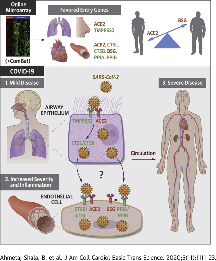
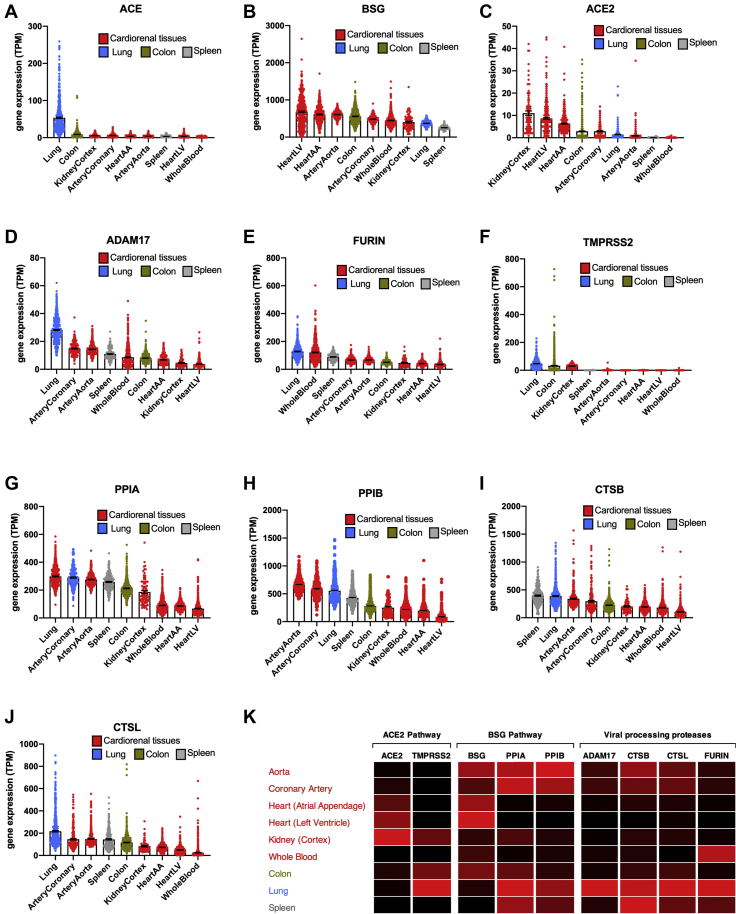
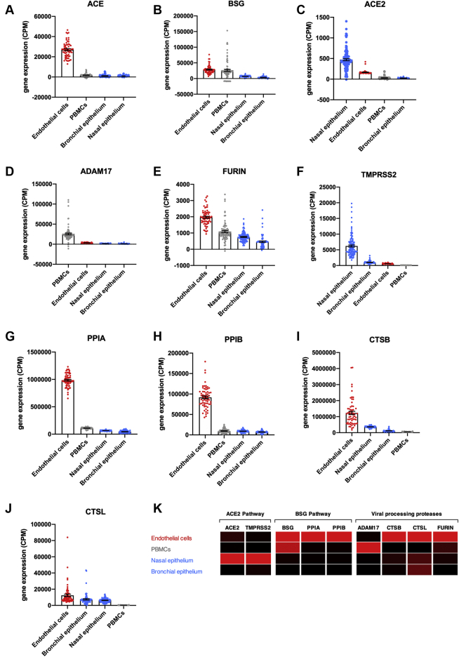
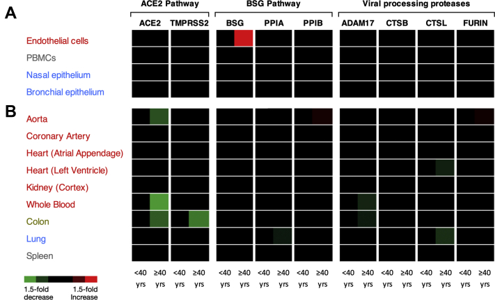
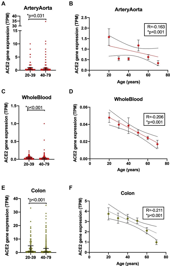
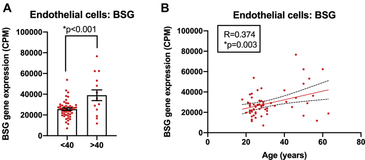
Vascular and cardiovascular inflammation and thrombosis occur in patients with severe coronavirus disease-2019 (COVID-19). Advancing age is the most significant risk factor for severe COVID-19. Using transcriptomic databases, the authors found that: 1) cardiovascular tissues and endothelial cells express putative genes for severe acute respiratory syndrome coronavirus-2 infection, including angiotensin-converting enzyme 2 (ACE2) and basigin (BSG); 2) severe acute respiratory syndrome coronavirus-2 receptor pathways ACE2/transmembrane serine protease 2 and BSG/peptidylprolyl isomerase B(A) polarize to lung/epithelium and vessel/endothelium, respectively; 3) expression of host genes is relatively stable with age; and 4) notable exceptions are ACE2, which decreases with age in some tissues, and BSG, which increases with age in endothelial cells, suggesting that BSG expression in the vasculature may explain the heightened risk for severe disease with age.

Keywords: ACE2, angiotensin converting enzyme 2; ADAM17, ADAM metallopeptidase domain 17; BSG, basigin; COVID-19; COVID-19, coronavirus disease-2019; CTSB, cathepsin B; CTSL, cathepsin L; GTEx, Genotype-Tissue Expression; PBMC, peripheral blood mononuclear cells; PPIA, peptidylprolyl isomerase A; PPIB, peptidylprolyl isomerase B; SARS-CoV-2, severe acute respiratory syndrome-coronavirus-2; TMPRSS2, transmembrane serine protease 2; age; cardiovascular; endothelial cells.


Conflict of interest statement

This work was funded by the Wellcome Trust/Imperial College Institutional Support Fellowship for Dr. Ahmetaj-Shala. Dr. Kirkby is a recipient of an Intermediate Research Fellowship from the British Heart Foundation (FS/16/1/31699). Dr. Vaja is a recipient of a Clinical Training Fellowship from the British Heart Foundation (FS/19/6/34129). Drs. Kirkby and Mitchell are holders of a program grant from the British Heart Foundation (RG/18/4/33541). All other authors have reported that they have no relationships relevant to the contents of this paper to disclose.

Figures

None
Graphical abstract
Figure 1
Figure 1
Expression of SARS-CoV-2 Entrance and Processing Genes in Different Cardiorenal and Noncardiorenal Tissues Standardized expression levels for the genes ACE(A), BSG(B), ACE2(C), ADAM17(D), FURIN(E), TMPRSS2(F), PPIA(G), PPIB(H), CTSB(I), and CTSL(J) were obtained from Genotype-Tissue Expression from cardiorenal tissues (aorta, coronary artery, atrial appendage [AA], left ventricle [LV], kidney cortex, and whole blood; red columns) and other tissues (lung, blue columns; spleen, gray columns; and colon, green columns). Data for each tissue corrected for batch effects using ComBat-seq and expressed as individual points and mean ± SEM. Tissues are ranked in order of expression for each gene. A heat map showing expression of angiotensin-converting enzyme 2 (ACE2) and basigin (BSG) pathways and associated viral processing proteases in each tissue was generated (K). Data are colored by gene, whereby black is the lowest expressing tissue and red is the highest expressing tissue. SARS-CoV-2 = severe acute respiratory syndrome coronavirus-2.
Figure 2
Figure 2
Expression of SARS-CoV-2 Entrance and Processing Genes in Blood Outgrowth Endothelial Cells, PBMCs, and Epithelial Cells (Nasal and Bronchial) Standardized expression levels for the genes ACE(A), BSG(B), ACE2(C), ADAM17(D), FURIN(E), TMPRSS2(F), PPIA(G), PPIB(H), CTSB(I), and CTSL(J) were obtained from online databases from human blood outgrowth endothelial cells (endothelial cells; red columns), peripheral blood mononuclear cells (PBMCs) (gray columns), and epithelial cells (nasal and bronchial; blue columns). The data were aligned and analyzed using Partek Flow and corrected for batch effects using ComBat-seq and expressed as individual data points and mean ± SEM. Cells were ranked in order of expression each gene. A heat map showing expression of ACE2 and BSG pathways and viral processing proteases in each cell type was generated (K). Data are colored by gene, whereby black is the lowest expressing cell type and red is the highest expressing cell type. Abbreviations as in Figure 1.
Figure 3
Figure 3
Heat Map Representing Expression of SARS-CoV-2 Entrance and Processing Genes Significantly Altered by Age Heat maps were generated for the expression of ACE, ACE2, BSG, ADAM17, CTSB, CTSL, FURIN, PPIA, PPIB, and TMPRSS2 in cells (A) and organs (B). Data were analyzed on the basis of 2 adult age groups; <40 or >40 years. Data were analyzed using an unpaired Mann-Whitney U test or Student’s t-test depending on normality. Significant data (p < 0.05) are shown as either increased (red) or decreased (green) expression; black corresponds to no significant change.
Figure 4
Figure 4
The Effect of Age on ACE2 Expression in Aorta, Whole Blood, and Colon ACE2 levels in aorta (A,B), whole blood (C,D), and colon (E,F) were analyzed on the basis of adults <40 versus >40 years of age (A,C,E). Data were analyzed using unpaired Mann-Whitney U test (A,C,E) or Spearman correlation test (B,D,F). Data are expressed as individual points and median ± interquartile range (A,C,E) or mean ± SEM (B,D,F); significance was accepted for ∗p < 0.05.
Figure 5
Figure 5
The Effect of Age on BSG Expression in Blood Outgrowth Endothelial Cells BSG levels in blood outgrowth endothelial cells (endothelial cells) were analyzed in adults <40 years of age versus ≥40 years of age using an unpaired Student’s t-test (A) and correlations with age determined using Pearson correlation analysis (B). Data are expressed as individual data points and mean ± SEM; significance was accepted at ∗p < 0.05.

Comment in

References

    1. Hoffmann M., Kleine-Weber H., Schroeder S. SARS-CoV-2 cell entry depends on ACE2 and TMPRSS2 and is blocked by a clinically proven protease inhibitor. Cell. 2020;181:271–280.e8. - PMC - PubMed
    1. Ulrich H., Pillat M.M. CD147 as a target for COVID-19 treatment: suggested effects of azithromycin and stem cell engagement. Stem Cell Rev Rep. 2020;16:434–440. - PMC - PubMed
    1. Wang K., Chen W., Zhou Y.-S. SARS-CoV-2 invades host cells via a novel route: CD147-spike protein. bioRxiv. https://www.biorxiv.org/content/10.1101/2020.03.14.988345v1 Available at: - DOI
    1. Coutard B., Valle C., de Lamballerie X., Canard B., Seidah N.G., Decroly E. The spike glycoprotein of the new coronavirus 2019-nCoV contains a furin-like cleavage site absent in CoV of the same clade. Antiviral Res. 2020;176:104742. - PMC - PubMed
    1. Shilts J., Wright G.J. No evidence for basigin/CD147 as a direct SARS-CoV-2 spike binding receptor. bioRxiv. https://www.biorxiv.org/content/10.1101/2020.07.25.221036v1 Available at: - DOI - PMC - PubMed