Recognizing differentiating clinical signs of CLN3 disease (Batten disease) at presentation
- PMID: 33073538
- PMCID: PMC8359263
- DOI: 10.1111/aos.14630
Recognizing differentiating clinical signs of CLN3 disease (Batten disease) at presentation
Abstract
Purpose: To help differentiate CLN3 (Batten) disease, a devastating childhood metabolic disorder, from the similarly presenting early-onset Stargardt disease (STGD1). Early clinical identification of children with CLN3 disease is essential for adequate referral, counselling and rehabilitation.
Methods: Medical chart review of 38 children who were referred to a specialized ophthalmological centre because of rapid vision loss. The patients were subsequently diagnosed with either CLN3 disease (18 patients) or early-onset STGD1 (20 patients).
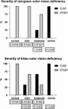
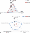
Results: Both children who were later diagnosed with CLN3 disease, as children who were later diagnosed with early-onset STGD1, initially presented with visual acuity (VA) loss due to macular dystrophy at 5-10 years of age. VA in CLN3 disease decreased significantly faster than in STGD1 (p = 0.01). Colour vision was often already severely affected in CLN3 disease while unaffected or only mildly affected in STGD1. Optic disc pallor on fundoscopy and an abnormal nerve fibre layer on optical coherence tomography were common in CLN3 disease compared to generally unaffected in STGD1. In CLN3 disease, dark-adapted (DA) full-field electroretinogram (ERG) responses were either absent or electronegative. In early-onset STGD1, DA ERG responses were generally unaffected. None of the STGD1 patients had an electronegative ERG.
Conclusion: Already upon presentation at the ophthalmologist, the retina in CLN3 disease is more extensively and more severely affected compared to the retina in early-onset STGD1. This results in more rapid VA loss, severe colour vision abnormalities and abnormal DA ERG responses as the main differentiating early clinical features of CLN3 disease.
Keywords: Batten disease; CLN3 disease; childhood retinal dystrophy; deep phenotyping; early recognition; early-onset STGD1.
© 2020 The Authors. Acta Ophthalmologica published by John Wiley & Sons Ltd on behalf of Acta Ophthalmologica Scandinavica Foundation.
Figures


References
-
- Audo I, Robson AG, Holder GE & Moore AT (2008): The negative ERG clinical phenotypes and disease mechanisms of inner retinal dysfunction. Surv Ophthalmol 53: 16–40. - PubMed
-
- Bax NM, Lambertus S, Cremers FPM, Klevering BJ & Hoyng CB (2019): The absence of fundus abnormalities in Stargardt disease. Graefes Arch Clin Exp Ophthalmol 257: 1147–1157. - PubMed
-
- Bohra LI, Weizer JS, Lee AG & Lewis RA (2000): Vision loss as the presenting sign in juvenile neuronal ceroid lipofuscinosis. J Neuroophthalmol 20: 111–115. - PubMed
-
- Brouwer AH, de Wit GC, Ten Dam NH, Wijnhoven R, van Genderen MM & de Boer JH (2019): Prolonged cone b‐wave on electroretinography is associated with severity of inflammation in non‐infectious uveitis. Am J Ophthalmol 207: 121–129. - PubMed
MeSH terms
Substances
Grants and funding
LinkOut - more resources
Full Text Sources

