. 2020 Nov;9(22):e2001657.
doi: 10.1002/adhm.202001657. Epub 2020 Oct 19.

Hybrid Bioprinting of Zonally Stratified Human Articular Cartilage Using Scaffold-Free Tissue Strands as Building Blocks

Affiliations

Hybrid Bioprinting of Zonally Stratified Human Articular Cartilage Using Scaffold-Free Tissue Strands as Building Blocks

Yang Wu et al. Adv Healthc Mater. 2020 Nov.

Abstract

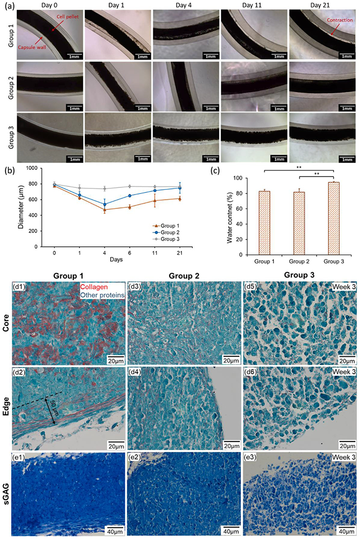
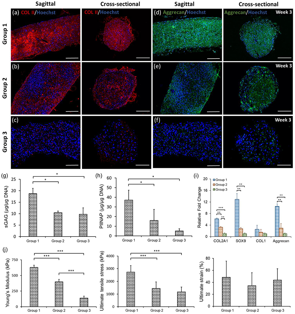
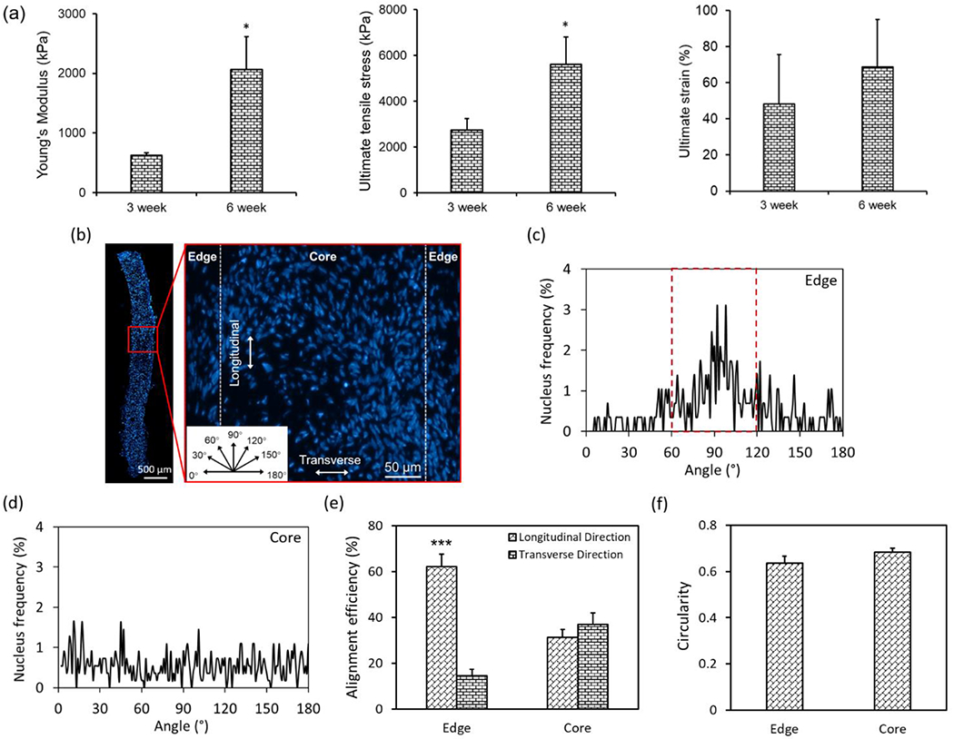
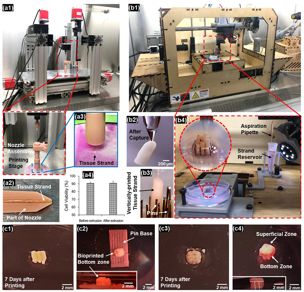
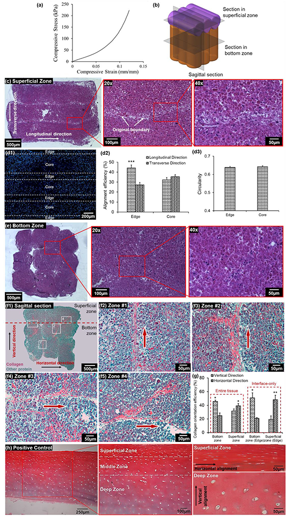
The heterogeneous and anisotropic articular cartilage is generally studied as a layered structure of "zones" with unique composition and architecture, which is difficult to recapitulate using current approaches. A novel hybrid bioprinting strategy is presented here to generate zonally stratified cartilage. Scaffold-free tissue strands (TSs) are made of human adipose-derived stem cells (ADSCs) or predifferentiated ADSCs. Cartilage TSs with predifferentiated ADSCs exhibit improved mechanical properties and upregulated expression of cartilage-specific markers at both transcription and protein levels as compared to TSs with ADSCs being differentiated in the form of strands and TSs of nontransfected ADSCs. Using the novel hybrid approach integrating new aspiration-assisted and extrusion-based bioprinting techniques, the bioprinting of zonally stratified cartilage with vertically aligned TSs at the bottom zone and horizontally aligned TSs at the superficial zone is demonstrated, in which collagen fibers are aligned with designated orientation in each zone imitating the anatomical regions and matrix orientation of native articular cartilage. In addition, mechanical testing study reveals a compression modulus of ≈1.1 MPa, which is similar to that of human articular cartilage. The prominent findings highlight the potential of this novel bioprinting approach for building biologically, mechanically, and histologically relevant cartilage for tissue engineering purposes.

Keywords: adipose-derived stem cells; biofabrication; scaffold-free bioprinting; zonally stratified articular cartilage.


Figures

Figure 1.
Figure 1.
Schematic overview of fabrication of scaffold-free zonally-stratified articular cartilage: human ADSCs were expanded and differentiated under chondrogenic induction followed by centrifuging them to obtain dense pellet. Cells were then microinjected into alginate capsules to facilitate cell aggregation followed by de-crosslinking of alginate capsules to obtain TSs. Zonally-stratified cartilage was comprised of the vertically aligned bottom zone, which was fabricated using an aspiration-assisted bioprinting (AAB) technique with the assistance of a pin array and the superficial zone, which was bioprinted using an extrusion-based bioprinting technique. After tissue fusion, the superficial zone was transferred onto the bottom zone and assembled via tissue fusion.
Figure 2.
Figure 2.
(a) Morphology and (b) diameter change of TSs in alginate capsules cultured for 3 weeks. (c) Water content of different groups of TSs (n=3, **p < 0.01). (d) Images of histological sections stained for Picrosirius Red/Fast Green demonstrating collagen content at the core and edges of TSs of different groups after the 3-week culture. (e) sGAG content stained by Toluidine Blue in different groups after the 3-week culture.
Figure 3.
Figure 3.
Expression of cartilage-specific markers, quantification of protein and gene expression, and mechanical properties in different groups of TSs. (a-c) COL-II and (d-f) Aggrecan immunostaining in the sagittal and cross-sectional views of TSs in different groups after the 3-week culture. (g) sGAG content measurement by DMMB assay, which was normalized to DNA amount (n=3, *p < 0.05). (h) COL-II synthesis from TSs with normalization to DNA amount (n=3, *p < 0.05). (i) Real-time PCR analysis of TSs demonstrating expression of cartilage-specific genes including COL2A1, Sox9, COL1 and Aggrecan in different groups (n=3, **p < 0.01, and ***p < 0.001). (j) Mechanical analysis of three types of TSs over the 3-week incubation period, including Young’s modulus, ultimate tensile stress and failure strain (n=3, ***p < 0.001).
Figure 4.
Figure 4.
Quantification of mechanical properties and cellular alignment and orientation within TSs in Group 1. (a) Comparison of mechanical properties of TSs in Group 1 after 3 and 6 weeks of culture (n=3, *p < 0.05). (b) A fluorescent image showing nuclei distribution throughout the histological section of a TS in Group 1 after 3 weeks of culture, which was divided into two regions (edge and core) based on the distance to edges (insert: angular orientation, which was used to define the nucleus angle). (c-d) Analysis of nucleus angle frequency at two regions. (e) Quantitative analysis of nucleus alignment efficiency and (f) circularity (n=4; ***p < 0.001).
Figure 5.
Figure 5.
Hybrid bioprinting of scaffold-free zonally-stratified articular cartilage: (a) bioprinting of the superficial zone using the MABP (a1) with a detachable nozzle assembly (a2). Loaded TSs were extruded with the movement of nozzle (a3). Extrusion process did not result in any significant changes in cell viability (a4). (b) Bioprinting of the bottom zone using the AAB technique (b1). The pick-and-place procedure (b2) was performed to place the TSs into the pin array repetitively (b3) to obtain a well-defined 3×3 arrangement of TSs (b4). (c) The bioprinted superficial zone (c1) was immobilized by alginate hydrogel and cultured for a week to facilitate tissue fusion. The bioprinted bottom zone was also immobilized by alginate hydrogel, followed by pins being removed (c2). The construct was then detached from the pin base (c3) and cultured for a week to facilitate tissue fusion. The bioprinted superficial zone was transferred onto the bottom zone and cultured for another week to generate the whole cartilage with two distinct zones (c4).
Figure 6.
Figure 6.
Characterization of fabricated zonally-stratified cartilage. (a) Stress-strain curves of the compression test on the bioprinted cartilage. (b) A schematic diagram showing positions of transverse and sagittal sectioning in the bioprinted articular cartilage. (c) H&E staining of the superficial zone with different magnifications demonstrating the fusion of TSs and cellular morphology. (d) A fluorescent image of nuclei on a section of the superficial zone, which was divided into two zones (edge and core) on each TS domain (d1), and the analysis of nuclei alignment (d2) and elongation (d3) of these regions (n=3, ***p < 0.001). (e) H&E straining of the bottom zone with different magnifications demonstrating the fusion of TSs and chondrocyte morphology. (f) Sagittal sections of bioprinted cartilage with Picrosirius Red/Fast Green straining demonstrating the stratified arrangement and collagen organization in (f1) whole cartilage and (f2-f5) at the boundary regions, which were labelled in (f1). (g) Quantitative measurement of collagen alignment efficiency at the superficial and bottom zones of the entire tissue, as well as at the interfaces of TSs (n=3, *p < 0.05 and **p < 0.01). (h) Picrosirius Red/Fast Green straining on a section of human cartilage as a positive control showing the zonally-stratified arrangement of collagen.

References

    1. Curl WW, Krome J, Gordon ES, Rushing J, Smith BP, Poehling GG, Arthroscopy 1997, 13, 456. - PubMed
    1. Farr J, Cole B, Dhawan A, Kercher J, Sherman S, Clin. Orthop. Relat. Res 2011, 469, 2696. - PMC - PubMed
    1. Zhang L, Hu J, Athanasiou KA, Crit. Rev. Bioeng 2009, 37, 1. - PMC - PubMed
    1. Laurencin CT, Ambrosio A, Borden M, Cooper J Jr, Annu. Rev. Biomed. Eng 1999, 1, 19. - PubMed
    1. Temenoff JS, Mikos AG, Biomaterials 2000, 21, 431. - PubMed

Publication types