. 2020 Nov;166(11):1038-1046.
doi: 10.1099/mic.0.000975. Epub 2020 Oct 5.

Siderophore piracy enhances Vibrio cholerae environmental survival and pathogenesis

Affiliations

Siderophore piracy enhances Vibrio cholerae environmental survival and pathogenesis

Hyuntae Byun et al. Microbiology (Reading). 2020 Nov.

Abstract

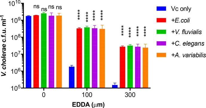
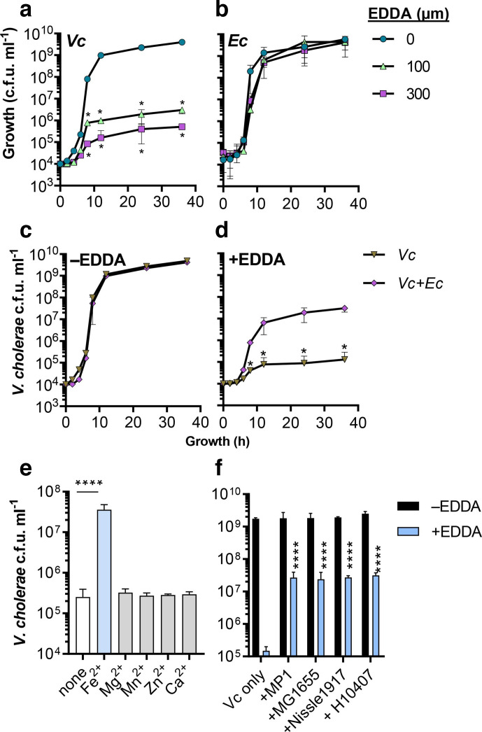
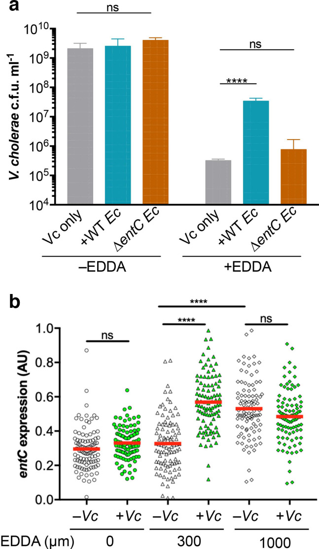
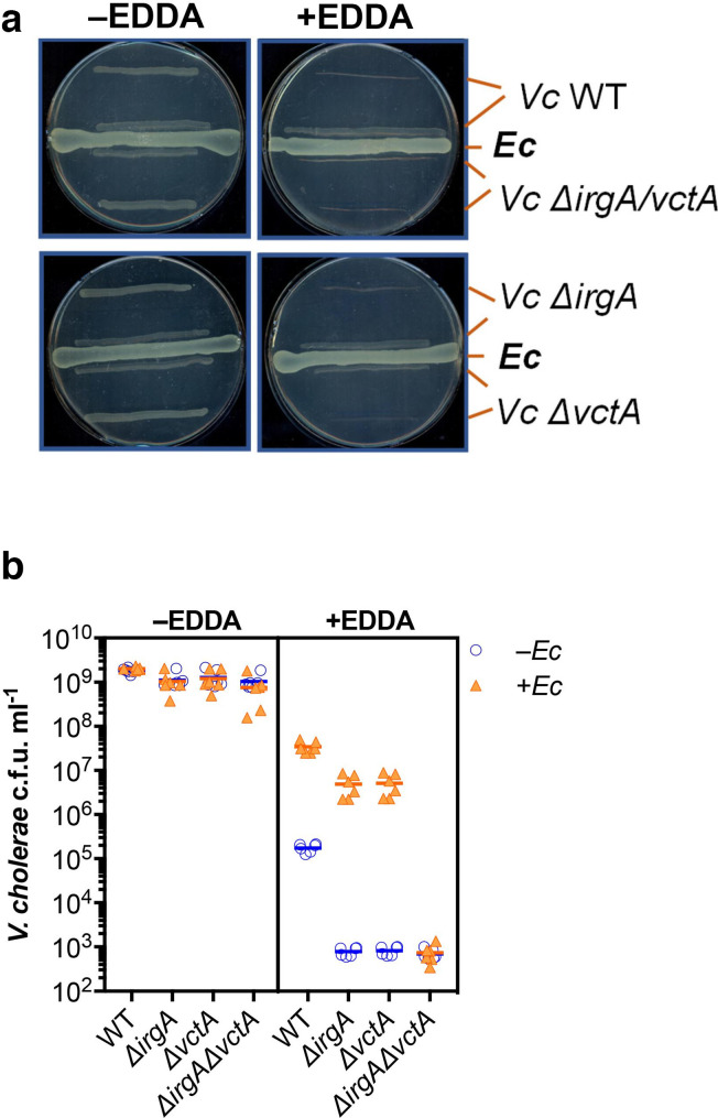
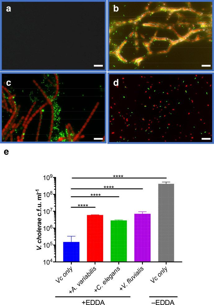
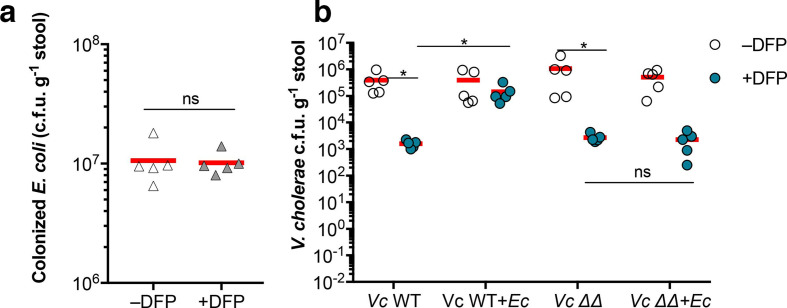
Vibrio cholerae, the aetiological agent of cholera, possesses multiple iron acquisition systems, including those for the transport of siderophores. How these systems benefit V. cholerae in low-iron, polymicrobial communities in environmental settings or during infection remains poorly understood. Here, we demonstrate that in iron-limiting conditions, co-culture of V. cholerae with a number of individual siderophore-producing microbes significantly promoted V. cholerae growth in vitro. We further show that in the host environment with low iron, V. cholerae colonizes better in adult mice in the presence of the siderophore-producing commensal Escherichia coli. Taken together, our results suggest that in aquatic reservoirs or during infection, V. cholerae may overcome environmental and host iron restriction by hijacking siderophores from other microbes.

Keywords: Anabaena variabilis; Cunninghamella elegans; Escherichia coli; Vibrio cholerae; Vibrio fluvialis; colonization; enterobactin; iron.


Conflict of interest statement

The authors declare that there are no conflicts of interest.

Figures

Fig. 1.
Fig. 1.
The growth of V. cholerae without or with siderophore-producing microbes under iron-limited conditions. V. cholerae was added to minimal medium containing the strain indicated and with glucose (for E. coli, C. elegans and V. fluvialis ) and fructose (for A. variabilis ) as carbon sources. Different concentrations of EDDA were included as indicated. The cultures were incubated at 37 °C with aeration for 18 h, and viable cells of V. cholerae were determined by serial dilution and plating on selective LB agar plates. The mean of three independent assays is shown and error bars represent the standard deviation. ****, P <0.0001 (two-way ANOVA, compared to V. cholerae -only samples); ns, no significance.
Fig. 2.
Fig. 2.
Effects of E. coli on V. cholerae growth in iron-limited conditions. (a, b) Singular growth of V. cholerae (a) and E. coli (b) under iron-limited conditions. One hundred thousand V. cholerae or E. coli were inoculated into 1 ml minimal medium with different concentrations of EDDA. Bacterial c.f.u. was determined at the time points indicated. The mean of three independent assays is shown, and error bars represent the standard deviation. *, P <0.05 (two-way ANOVA, compared to 0 EDDA data points). (c, d) V. cholerae and E. coli co-cultures. One hundred thousand V. cholerae and 106 E. coli were inoculated into the minimal medium without (c) or with (d) 300 µm EDDA. V. cholerae c.f.u. was determined at the time points indicated. The mean of three independent assays is shown, and error bars represent the standard deviation. (e) The effect of divalent metals on V. cholerae growth. Sixty micrometres of various metals were included in the minimal medium containing 300 µm EDDA. V. cholerae c.f.u. was then determined after 18 h incubation. The mean of three independent assays is shown and error bars represent the standard deviation. *, P <0.05. (f) V. cholerae co-cultures with different E. coli strains. V. cholerae and E. coli strains were inoculated at a 1 :10 ratio and incubated in the minimal medium without or with 300 µm EDDA. The cultures were incubated at 37 °C with aeration for 18 h, and the c.f.u. of V. cholerae was determined. The mean of three independent assays is shown and error bars represent the standard deviation. ****, P <0.0001 (two-way ANOVA, compared to V. cholerae -only samples).
Fig. 3.
Fig. 3.
E. coli -produced enterobactin utilization by V. cholerae . (a) The requirement of the entC gene. One hundred thousand V. cholerae either alone or in co-culture with either 106 MP1 or MP1 ΔentC were inoculated into the minimal medium with or without 300 µm EDDA. V. cholerae c.f.u. was determined after 16 h incubation. The mean of four independent assays is shown and error bars represent the standard deviation. ***, P <0.0005 (Student’s t-test); ns, no significance. (b) entC expression. One million E. coli entC-gfp reporter strains were inoculated into 1 ml minimal medium containing different indicated concentrations of EDDA with and without V. cholerae . Tetracycline (2 µg ml−1) was also included in the medium to induce Ptet-mcherry. The cultures were growth at 37 °C with aeration for 16 h. Bacterial cells were examined using a Nikon NiU fluorescence microscope with default settings. The GFP intensity of each cell was quantified by using the Nis-elements basic research microscope imaging software and normalized against mCherry signals. For each condition, 100 cells from 3 independent samples were analysed. ****, P <0.0001 (two-way ANOVA). ns, no significance.
Fig. 4.
Fig. 4.
The role of V. cholerae receptors in co-culture. (a) Cross-feeding experiments. E. coli MP1 and V. cholerae wild-type, ΔirgA, ΔvctA and ΔirgAΔvctA were streaked on LB agar without and with 3 mM EDDA. The plates were incubated at 37 °C for 24 h. (b) V. cholerae wild-type, ΔirgA, ΔvctA and ΔirgAΔvctA co-cultures with E. coli with and without EDDA. V. cholerae and E. coli strains were inoculated at a 1 :10 ratio and incubated in the minimal medium containing 300 µm of EDDA. The mean of six independent assays is shown and error bars represent the standard deviation. ****, P <0.0001 (Student’s t-test); ns, no significance.
Fig. 5.
Fig. 5.
V. cholerae growth in seawater-based growth medium with siderophore-producing microbes. GFP-labelled V. cholerae alone or with other indicated siderophore-producing microbes were grown in EDDA-treated seawater medium for various times at 22 ˚C. The samples were examined using a Nikon NiU fluorescence microscope according to the manufacturer’s instructions. The images are a composite of mCherry and GFP filters. (a) V. cholerae alone. (b–d) V. cholerae with C. elegans (b), A. variabilis (c) and V. fluvialis containing an mCherry-expressing plasmid (d). Scale bar, 10 µm. (e) V. cholerae growth in the presence of siderophore-producing marine organisms in iron-limiting seawater medium. The mean of three independent assays is shown and error bars represent the standard deviation. ****, P <0.0001 (Student’s t-test, compared to V. cholerae -only sample+EDDA).
Fig. 6.
Fig. 6.
Enterobactin utilization by V. cholerae in vivo. Mouse colonization. Six-week-old CD-1 mice without and with DFP were treated with streptomycin for 1 day. Approximately 109 cells of wild-type V. cholerae (lacZ+) and ΔvctAΔirgA mutants (lacZ )(ΔΔ) were mixed at a 1 :1 ratio without or with wild-type streptomycin- and tetracycline-resistant MP1 and inoculated intragastrically into mice. After 3 days of colonization, fresh faecal pellets were collected, homogenized, serial diluted and plated on selective LB agar plates to determine the colonized E. coli (a) and V. cholerae (b). Horizonal lines, average number of colonized bacteria from five mice. *, P <0.05 (Student’s t-test). ns, no significance.

Similar articles

Cited by

References

    1. Miethke M, Marahiel MA. Siderophore-based iron acquisition and pathogen control. Microbiol Mol Biol Rev. 2007;71:413–451. doi: 10.1128/MMBR.00012-07. - DOI - PMC - PubMed
    1. Hider RC, Kong X. Chemistry and biology of siderophores. Nat Prod Rep. 2010;27:637–657. doi: 10.1039/b906679a. - DOI - PubMed
    1. Schaible UE, Kaufmann SH. Iron and microbial infection. Nat Rev Microbiol. 2004;2:946–953. doi: 10.1038/nrmicro1046. - DOI - PubMed
    1. Skaar EP. The battle for iron between bacterial pathogens and their vertebrate hosts. PLoS Pathog. 2010;6:e1000949. doi: 10.1371/journal.ppat.1000949. - DOI - PMC - PubMed
    1. Cassat JE, Skaar EP. Iron in infection and immunity. Cell Host Microbe. 2013;13:509–519. doi: 10.1016/j.chom.2013.04.010. - DOI - PMC - PubMed

Publication types

MeSH terms

LinkOut - more resources