. 2021 Jan;64(1):115-125.
doi: 10.1165/rcmb.2020-0360OC.

Lung Myofibroblasts Promote Macrophage Profibrotic Activity through Lactate-induced Histone Lactylation

Affiliations

Lung Myofibroblasts Promote Macrophage Profibrotic Activity through Lactate-induced Histone Lactylation

Huachun Cui et al. Am J Respir Cell Mol Biol. 2021 Jan.

Abstract

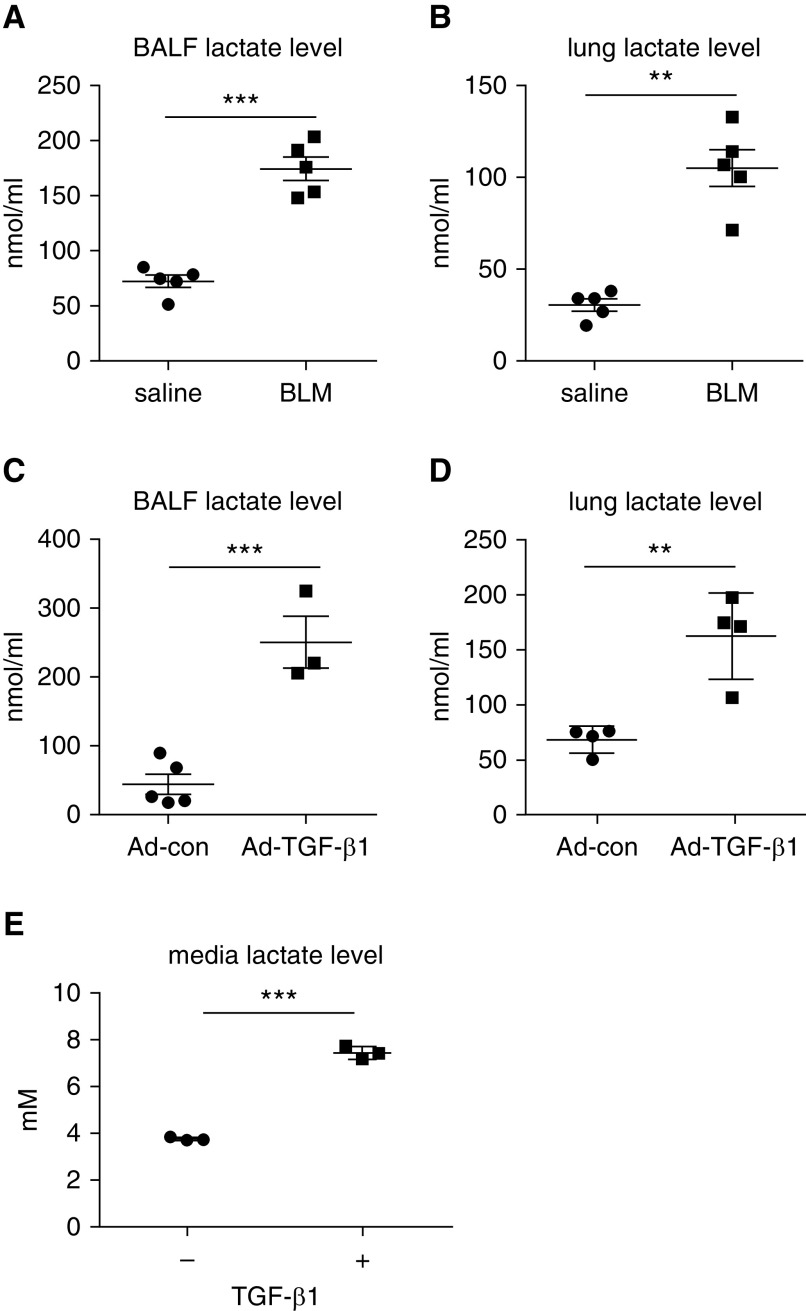
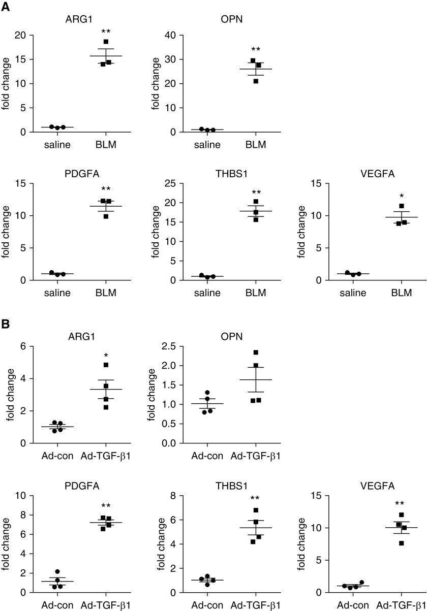
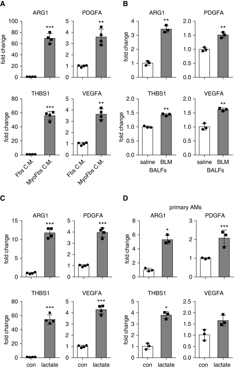
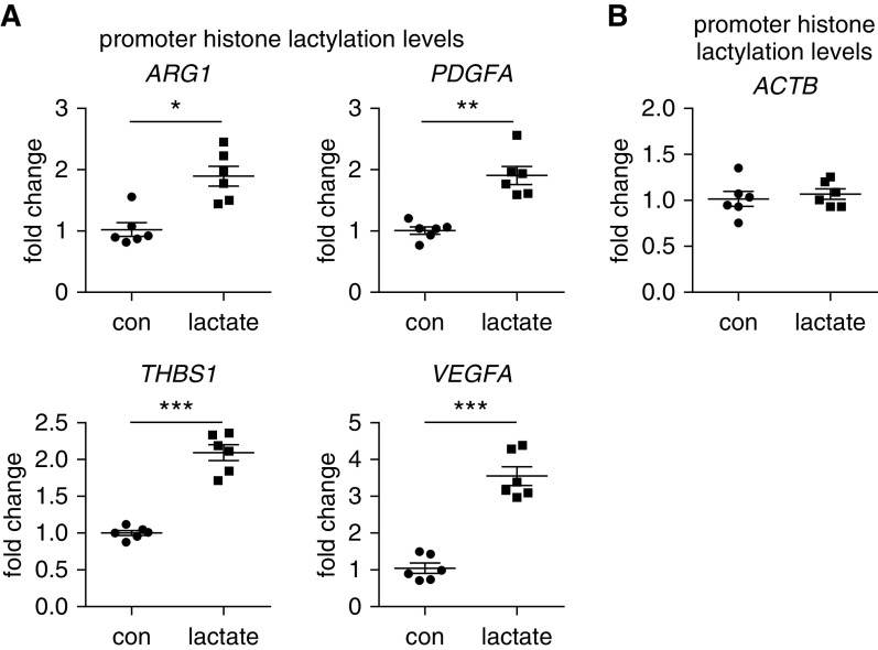
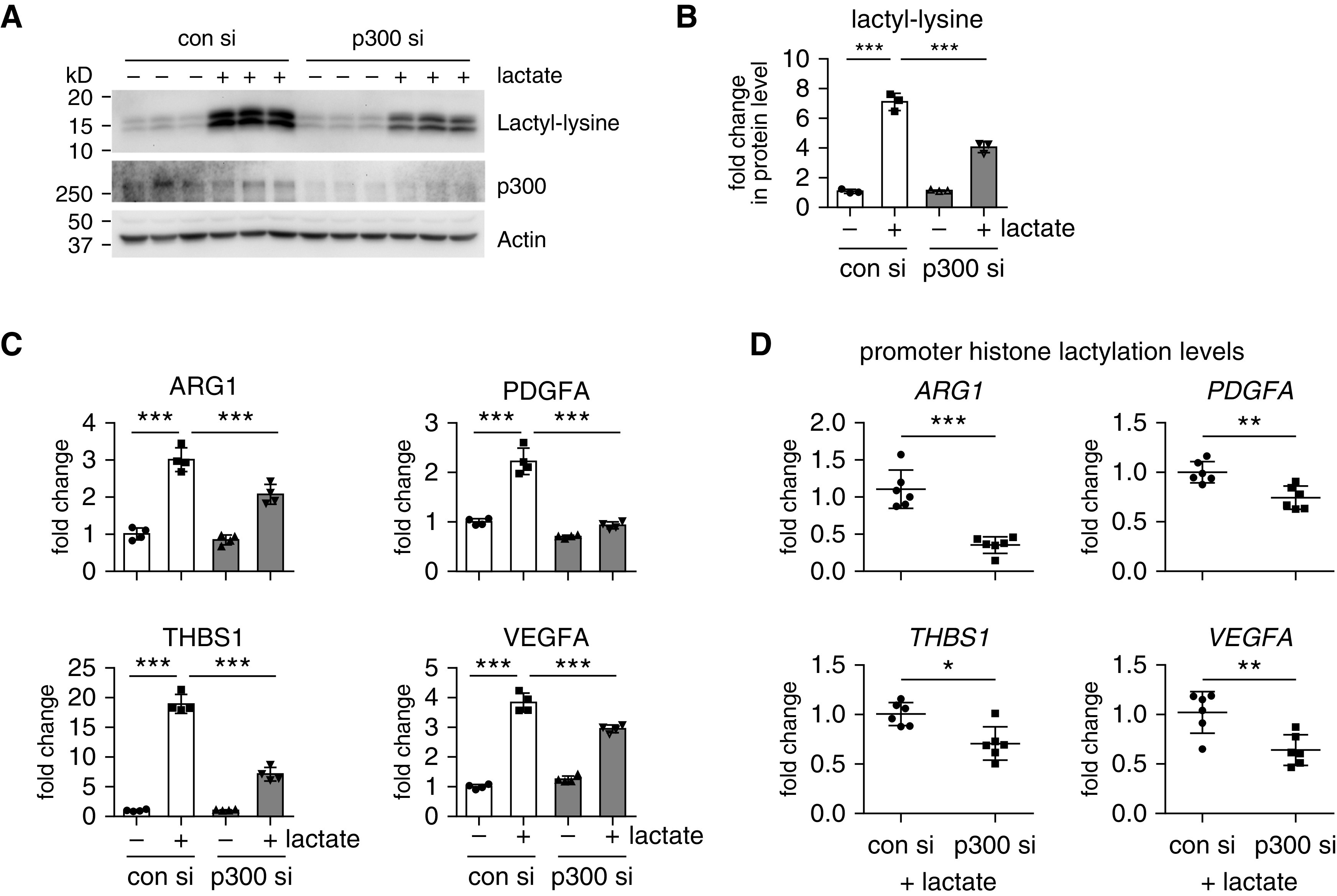
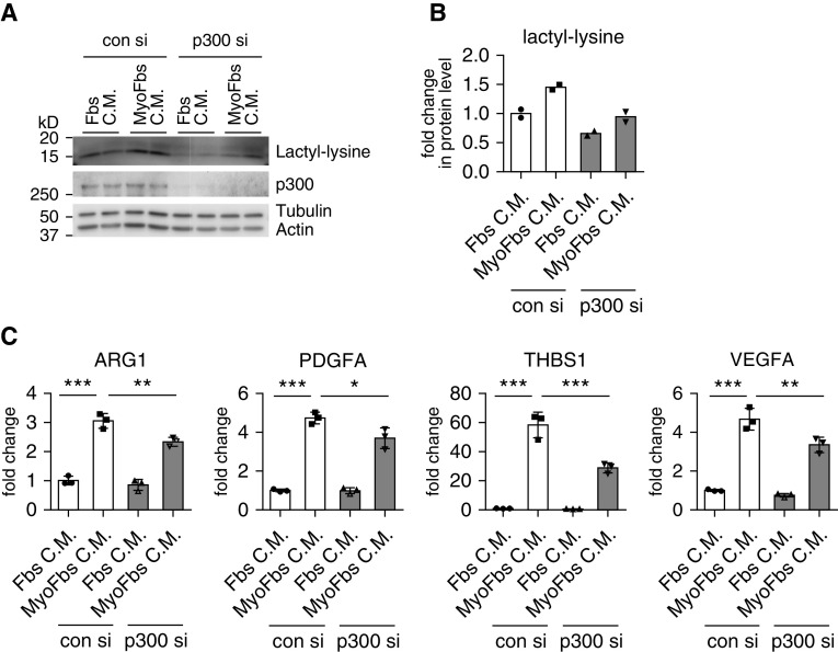
Augmented glycolysis due to metabolic reprogramming in lung myofibroblasts is critical to their profibrotic phenotype. The primary glycolysis byproduct, lactate, is also secreted into the extracellular milieu, together with which myofibroblasts and macrophages form a spatially restricted site usually described as fibrotic niche. Therefore, we hypothesized that myofibroblast glycolysis might have a non-cell autonomous effect through lactate regulating the pathogenic phenotype of alveolar macrophages. Here, we demonstrated that there was a markedly increased lactate in the conditioned media of TGF-β1 (transforming growth factor-β1)-induced lung myofibroblasts and in the BAL fluids (BALFs) from mice with TGF-β1- or bleomycin-induced lung fibrosis. Importantly, the media and BALFs promoted profibrotic mediator expression in macrophages. Mechanistically, lactate induced histone lactylation in the promoters of the profibrotic genes in macrophages, consistent with the upregulation of this epigenetic modification in these cells in the fibrotic lungs. The lactate inductions of the histone lactylation and profibrotic gene expression were mediated by p300, as evidenced by their diminished concentrations in p300-knockdown macrophages. Collectively, our study establishes that in addition to protein, lipid, and nucleic acid molecules, a metabolite can also mediate intercellular regulations in the setting of lung fibrosis. Our findings shed new light on the mechanism underlying the key contribution of myofibroblast glycolysis to the pathogenesis of lung fibrosis.

Keywords: histone lactylation; lactate; macrophage; myofibroblast; pulmonary fibrosis.


Figures

Figure 1.
Figure 1.
Increased glycolysis is characteristic of lung fibrosis. (A and B) Eight-week old C57BL/6 mice were i.t. instilled with saline or bleomycin (1.5 U/kg in 50 μl saline). Three weeks after treatment, mice were killed, and their BAL fluids (BALFs) and lungs were harvested. Lactate concentrations in the BALFs and lungs were determined (n = 5 mice for each group; mean ± SEM). **P < 0.01 and ***P < 0.001 by two-tailed Student’s t test. (C and D) C57BL/6 mice were i.t. instilled with control adenovirus or adenovirus expressing active TGF-β1 (transforming growth factor-β1) (1 × 1011 pfu/kg body weight). Two weeks after treatment, mice were killed, and lactate concentrations in the BALFs and lungs were determined (n = 3–5 mice for each group; mean ± SEM). **P < 0.01 and ***P < 0.001 by two-tailed Student’s t test. (E) Human lung fibroblast MRC5 cells were treated with or without TGF-β1 (2 ng/ml) for 48 hours. Lactate concentrations in the culture media were determined (n = 3; mean ± SD). ***P < 0.001 by two-tailed Student’s t test. Ad-con = control adenovirus; Ad-TGF-β1 = adenovirus expressing active TGF-β1; BLM = bleomycin; pfu = plaque-forming unit.
Figure 2.
Figure 2.
Alveolar macrophages in fibrotic lungs demonstrate profibrotic phenotype. (A) C57BL/6 mice were i.t. instilled with saline or BLM. Three weeks after treatment, BALFs were harvested, and alveolar macrophages were isolated. Total RNAs were purified, and concentrations of the indicated genes were determined by real-time PCR (n = 3 mice for each group; mean ± SEM). *P < 0.05 and **P < 0.01 by two-tailed Student’s t test. (B) C57BL/6 mice were i.t. instilled with Ad-con or Ad-TGF-β1. Two weeks after treatment, alveolar macrophages were isolated, and total RNAs were purified. Concentrations of the indicated genes were determined by real-time PCR (n = 4 mice for each group; mean ± SEM). *P < 0.05 and **P < 0.01 by two-tailed Student’s t test. ARG1 = arginase 1; OPN = osteopontin; PDGFA = platelet-derived growth factor A; THBS1 = thrombospondin 1; VEGFA = vascular endothelial growth factor A.
Figure 3.
Figure 3.
Lung myofibroblast–conditioned media, BALFs from mice with lung fibrosis, or lactate promote macrophage profibrotic phenotype. (A) Bone marrow–derived macrophages (BMDMs) were treated with conditioned media from normal fibroblasts or TGF-β1–induced myofibroblasts. Twenty-four hours after treatment, cells were harvested, and concentrations of the indicated genes were determined by real-time PCR (n = 4; mean ± SD). **P < 0.01 and ***P < 0.001 by two-tailed Student’s t test. (B) BMDMs were treated with BALFs from mice that were treated i.t. with saline or BLM for 3 weeks. Twenty-four hours after treatment, concentrations of the indicated genes were determined by real-time PCR. n = 3; mean ± SD. **P < 0.01 by two-tailed Student’s t test. (C) BMDMs were treated with or without 20 mM lactate for 24 hours. Cells were harvested, and concentrations of the indicated genes were determined by real-time PCR (n = 4; mean ± SD). ***P < 0.001 by two-tailed Student’s t test. (D) Primary alveolar macrophages) were isolated from BALFs of C57BL/6 mice and treated with 20 mM lactate for 24 hours. Concentrations of the indicated genes were determined by real-time PCR (n = 3; mean ± SD). *P < 0.05 and ***P < 0.001 by two-tailed Student’s t test. AM = alveolar macrophage; con = control; Fbs C.M. = fibroblast-conditioned media; MyoFbs C.M. = myofibroblast-conditioned media.
Figure 4.
Figure 4.
Lactate induces histone lactylation in macrophages. (A) BMDMs were treated with or without 20 mM lactate for 24 hours. The cells were lysed, and lactate contents in the cellular lysates were determined (n = 3; mean ± SD). ***P < 0.001 by two-tailed Student’s t test. (B) BMDMs were treated with or without 20 mM lactate as in A. Concentrations of the indicated proteins in whole cell lysates or HCl-extracted histones were determined by Western blotting. (C) BMDMs were treated with or without lactate. The entire blot was developed for lactyl-lysine. (D) BMDMs were treated with conditioned media from normal fibroblasts or TGF-β1–induced myofibroblasts. Twenty-four hours after treatment, cells were lysed, and lactate contents in the cellular lysates were determined (n = 3; mean ± SD). *P < 0.05 by two-tailed Student’s t test. (E) BMDMs were treated with conditioned media from normal fibroblasts or TGF-β1–induced myofibroblasts as in (D). Concentrations of the indicated proteins were determined by Western blotting. (F) C57BL/6 mice were i.t. instilled with saline or BLM. Three weeks after treatment, mice were killed, and lung slices were prepared. The expression of F4/80 and lactyl-histone were evaluated by immunofluorescence staining and fluorescence microscopy. (G) Slices of normal con and idiopathic pulmonary fibrosis lungs were prepared. The expression of CD68 and lactyl-histone were evaluated by immunofluorescence staining and fluorescence microscopy. (H and I) C57BL/6 mice were i.t. instilled with saline or BLM. Three weeks after treatment, BALFs were harvested, and alveolar macrophages were isolated and cultured on coverslips. The expression of F4/80 and lactyl-histone were evaluated by immunofluorescence staining and fluorescence microscopy (H). Single-cell fluorescence intensity was measured by ImageJ for ∼100 randomly selected cells from each group, and the average was calculated. ***P < 0.001 by two-tailed Student’s t test. The box-and-whisker plots depict the 25th and 75th percentiles and the median, minimum, and maximum values (I). (FH) Nuclei were counterstained with DAPI. Scale bars, 100 μm. AE = HCl-extracted; IPF = idiopathic pulmonary fibrosis; MFI = mean fluorescence intensity; WCE = whole cell lysates.
Figure 5.
Figure 5.
Lactate increases histone lactylation amounts at the promoters of the profibrotic genes in macrophages. BMDMs were treated with or without 20 mM lactate. Twenty-four hours after treatment, the cells were fixed with 1% formaldehyde and collected in radioimmunoprecipitation assay (RIPA) buffer for sonication. A chromatin IP assay was performed using anti–lactyl-lysine antibody. (A and B) The histone lactylation amounts within the proximal promoter regions of the indicated genes (A) and reference ACTB gene (B) were determined by real-time PCR (n = 6; mean ± SD). *P < 0.05, **P < 0.01, and ***P < 0.001 by two-tailed Student’s t test. ACTB = actin β.
Figure 6.
Figure 6.
p300 mediates lactate-induced histone lactylation and profibrotic mediator expression in macrophages. (A and B) BMDMs were transfected with 20 nM con siRNAs or p300 siRNAs. Twenty-four hours after transfection, the cells were treated with or without 20 nM lactate for another 24 hours. The histone lactylation amounts were determined by Western blotting (A) and densitometric analyses performed using ImageJ (B) (n = 3; mean ± SD). ***P < 0.001 by one-way ANOVA with Bonferroni’s post hoc test. (C) BMDMs were transfected and treated as in A and B. The concentrations of the indicated genes were determined by real-time PCR. ***P < 0.001 by one-way ANOVA with Bonferroni’s post hoc test. (D) BMDMs were transfected and treated as in AC. The histone lactylation amounts at the promoters of the indicated genes were determined by chromatin IP assay as in Figure 5 (n = 6; mean ± SD). *P < 0.05, **P < 0.01, and ***P < 0.001 by two-tailed Student’s t test. si = siRNAs.
Figure 7.
Figure 7.
Knockdown p300 attenuates the profibrotic phenotype induced by lung myofibroblast–conditioned media in macrophages. (A and B) BMDMs were transfected with 20 nM con si or p300 si. Twenty-four hours after transfection, the cells were treated with conditioned media from normal fibroblasts or TGF-β1–induced myofibroblasts for another 24 hours. The histone lactylation amounts were determined by Western blotting (A) and densitometric analyses performed using ImageJ (B). (C) BMDMs were transfected and treated as in A and B. The concentrations of the indicated genes were determined by real-time PCR. *P < 0.05, **P < 0.01, and ***P < 0.001 by one-way ANOVA with Bonferroni’s post hoc test.

Comment in

References

    1. Noble PW, Barkauskas CE, Jiang D. Pulmonary fibrosis: patterns and perpetrators. J Clin Invest. 2012;122:2756–2762. - PMC - PubMed
    1. Fernandez IE, Eickelberg O. New cellular and molecular mechanisms of lung injury and fibrosis in idiopathic pulmonary fibrosis. Lancet. 2012;380:680–688. - PubMed
    1. Mora AL, Rojas M, Pardo A, Selman M. Emerging therapies for idiopathic pulmonary fibrosis, a progressive age-related disease. Nat Rev Drug Discov. 2017;16:755–772. - PubMed
    1. Brownell R, Kaminski N, Woodruff PG, Bradford WZ, Richeldi L, Martinez FJ, et al. Precision medicine: the new frontier in idiopathic pulmonary fibrosis. Am J Respir Crit Care Med. 2016;193:1213–1218. - PMC - PubMed
    1. Hinz B, Phan SH, Thannickal VJ, Galli A, Bochaton-Piallat ML, Gabbiani G. The myofibroblast: one function, multiple origins. Am J Pathol. 2007;170:1807–1816. - PMC - PubMed

Publication types

MeSH terms