

Should Fetal Growth Charts Be References or Standards?
- PMID: 33074926
- PMCID: PMC7707154
- DOI: 10.1097/EDE.0000000000001275
Should Fetal Growth Charts Be References or Standards?
Abstract
Background: Fetal growth standards (prescriptive charts derived from low-risk pregnancies) are theoretically better tools to monitor fetal growth than conventional references. We examined how modifying chart inclusion criteria influenced the resulting curves.
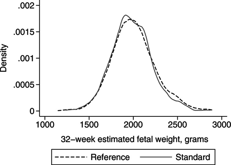
Methods: We summarized estimated fetal weight (EFW) distributions from a hospital's routine 32-week ultrasound in all nonanomalous singleton fetuses (reference) and in those without maternal-fetal conditions affecting fetal growth (standard). We calculated EFWs for the 3rd, 5th, 10th, and 50th percentiles, and the proportion of fetuses each chart classified as small for gestational age.
Results: Of 2309 fetuses in our reference, 690 (30%) met the standard's inclusion criteria. There were no meaningful differences between the EFW distributions of the reference and standard curves (50th percentile: 1989 g reference vs. 1968 g standard; 10th percentile: 1711 g reference vs. 1710 g standard), or the proportion of small for gestational age fetuses (both 9.9%).
Conclusions: In our study, there was little practical difference between a fetal growth reference and standard for detecting small infants.
Conflict of interest statement
The authors report no conflicts of interest.
Figures
References
-
- Hadlock FP, Harrist RB, Martinez-Poyer J. In utero analysis of fetal growth: a sonographic weight standard. Radiology. 1991;181:129–133. - PubMed
-
- Lubchenco LO, Hansman C, Dressler M, Boyd E. Intrauterine growth as estimated from liveborn birth-weight data at 24 to 42 weeks of gestation. Pediatrics. 1963;32:793–800. - PubMed
-
- McIntire DD, Bloom SL, Casey BM, Leveno KJ. Birth weight in relation to morbidity and mortality among newborn infants. N Engl J Med. 1999;340:1234–1238. - PubMed
-
- Ioannou C, Talbot K, Ohuma E, et al. Systematic review of methodology used in ultrasound studies aimed at creating charts of fetal size. BJOG. 2012;119:1425–1439. - PubMed
Publication types
MeSH terms
LinkOut - more resources
Full Text Sources
Miscellaneous
