. 2020 Dec:86:101793.
doi: 10.1016/j.compmedimag.2020.101793. Epub 2020 Sep 28.

The optimisation of deep neural networks for segmenting multiple knee joint tissues from MRIs

Affiliations

The optimisation of deep neural networks for segmenting multiple knee joint tissues from MRIs

Dimitri A Kessler et al. Comput Med Imaging Graph. 2020 Dec.

Abstract

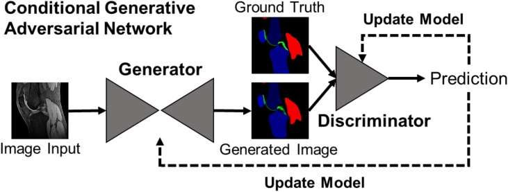
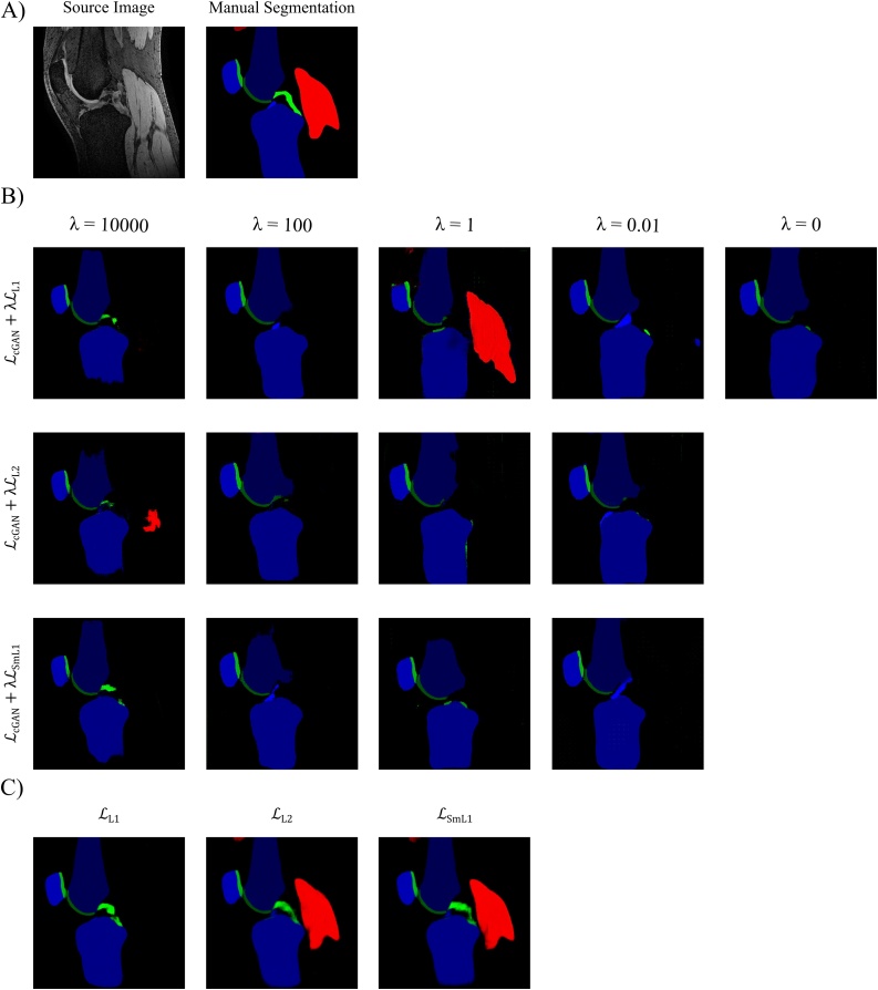
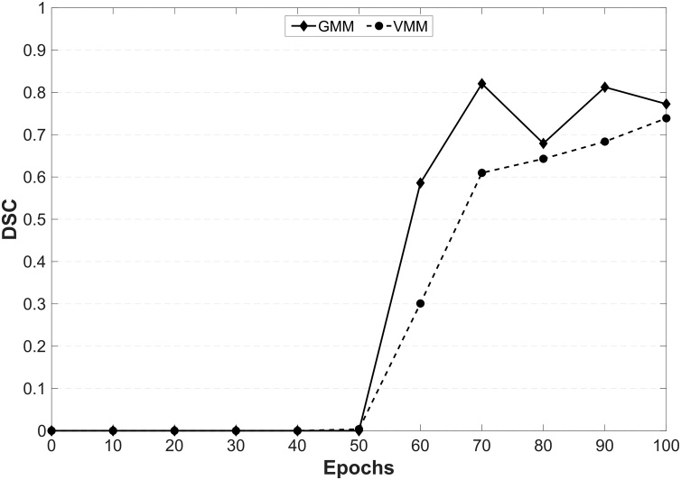
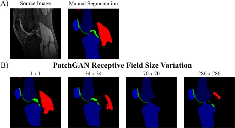
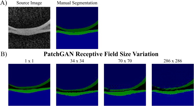
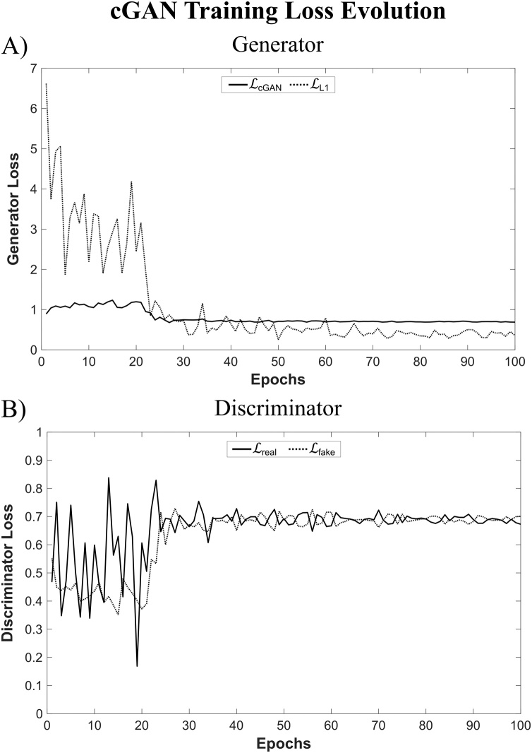
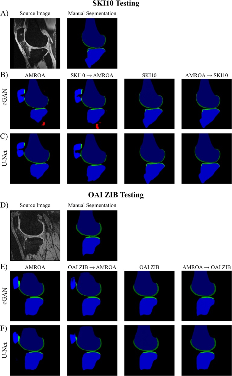
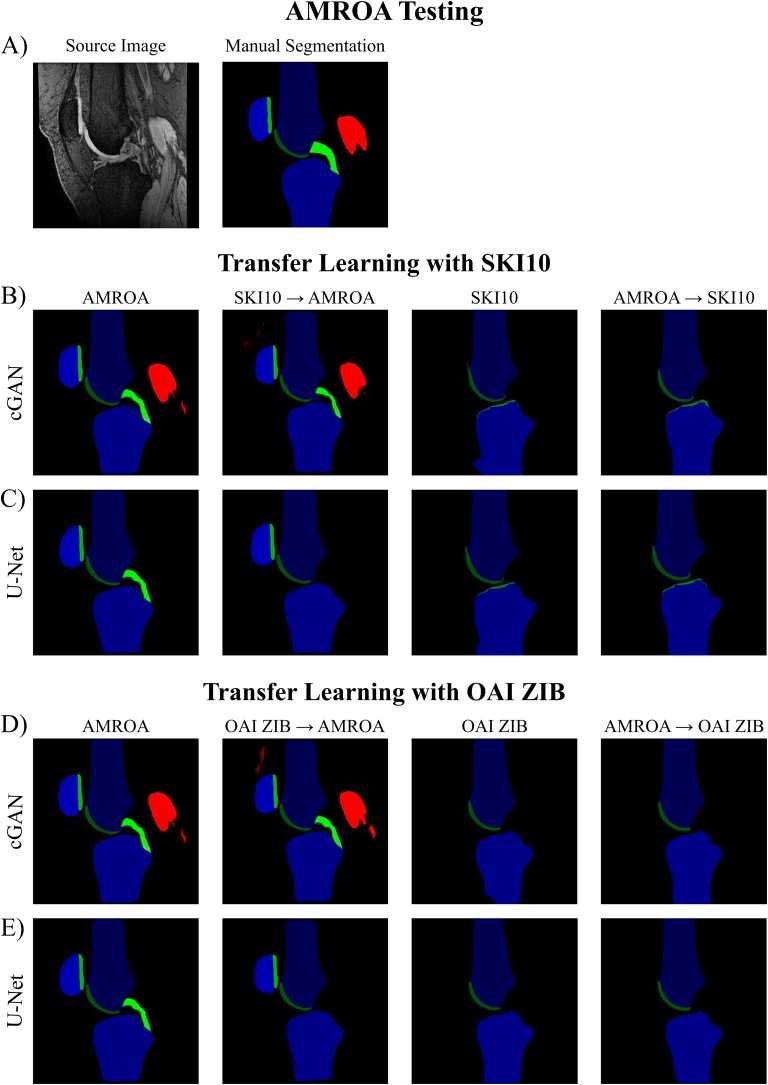
Automated semantic segmentation of multiple knee joint tissues is desirable to allow faster and more reliable analysis of large datasets and to enable further downstream processing e.g. automated diagnosis. In this work, we evaluate the use of conditional Generative Adversarial Networks (cGANs) as a robust and potentially improved method for semantic segmentation compared to other extensively used convolutional neural network, such as the U-Net. As cGANs have not yet been widely explored for semantic medical image segmentation, we analysed the effect of training with different objective functions and discriminator receptive field sizes on the segmentation performance of the cGAN. Additionally, we evaluated the possibility of using transfer learning to improve the segmentation accuracy. The networks were trained on i) the SKI10 dataset which comes from the MICCAI grand challenge "Segmentation of Knee Images 2010″, ii) the OAI ZIB dataset containing femoral and tibial bone and cartilage segmentations of the Osteoarthritis Initiative cohort and iii) a small locally acquired dataset (Advanced MRI of Osteoarthritis (AMROA) study) consisting of 3D fat-saturated spoiled gradient recalled-echo knee MRIs with manual segmentations of the femoral, tibial and patellar bone and cartilage, as well as the cruciate ligaments and selected peri-articular muscles. The Sørensen-Dice Similarity Coefficient (DSC), volumetric overlap error (VOE) and average surface distance (ASD) were calculated for segmentation performance evaluation. DSC ≥ 0.95 were achieved for all segmented bone structures, DSC ≥ 0.83 for cartilage and muscle tissues and DSC of ≈0.66 were achieved for cruciate ligament segmentations with both cGAN and U-Net on the in-house AMROA dataset. Reducing the receptive field size of the cGAN discriminator network improved the networks segmentation performance and resulted in segmentation accuracies equivalent to those of the U-Net. Pretraining not only increased segmentation accuracy of a few knee joint tissues of the fine-tuned dataset, but also increased the network's capacity to preserve segmentation capabilities for the pretrained dataset. cGAN machine learning can generate automated semantic maps of multiple tissues within the knee joint which could increase the accuracy and efficiency for evaluating joint health.

Keywords: Convolutional neural network (CNN); Generative adversarial network (GAN); Image segmentation; Magnetic resonance imaging (MRI); Musculoskeletal.


Conflict of interest statement

The authors report no declarations of interest.

Figures

Fig. 1
Fig. 1
Conditional GAN structure. The generator is a U-Net that progressively down-samples / encodes and then up-samples / decodes an input by a series of convolutional layers, with additional skip-connections between each major layer. The generated, ’fake’ segmentation image is then fed together with the ground truth segmentation image into a discriminator network (PatchGAN (Isola et al., 2017)) that gives its prediction of whether the generated image is a ‘real’ representation of the ground truth image, or not. A detailed description of the network architecture can be found in the Appendix.
Fig. 2
Fig. 2
Results of Network Objective Function. Qualitative results of B) training a cGAN with different objective functions by combining the cGAN loss with different pixel-wise error losses with varying weightings and C) training a U-Net with different pixel-wise error losses.
Fig. 3
Fig. 3
Results of testing on noise only images. Assessing the segmentation performance of a cGAN trained with LcGAN+λLL1(λ=100) loss objective and a U-Net trained with LL1 objective and tested on noise only images. Training was performed on the AMROA training dataset without noise only images. A) and B) are two example results of testing the models on noise only source images and comparing to ground truth segmentation maps.
Fig. 4
Fig. 4
Results of Altering the Loss Objective during Training. Assessing the influence of varying the objective function halfway during cGAN and U-Net training on their segmentation performance with comparison to the respective cGANs and U-Nets trained with constant loss function.
Fig. 5
Fig. 5
Influence of altering the loss objective during cGAN training on the segmentation performance of the medial gastrocnemius and vastus muscles. The cGAN was trained with a LcGAN+λLL2 loss objective for 50 epochs followed by a further 50 epochs training with LcGAN+λLL1. Abbreviations: VMM - vastus medialis muscle, GMM – medial head of gastrocnemius muscle, DSC – Dice Similarity Coefficient
Fig. 6
Fig. 6
Results of PatchGAN Receptive Field Size. Assessing the influence of varying the discriminator receptive field size on segmentation performance of cGAN when trained and tested on the AMROA dataset.
Fig. 7
Fig. 7
Image Artefact due to the choice of PatchGAN Receptive Field Size. Influence of discriminator receptive field size on checkerboard artefact emergence of a cGAN trained and tested on the AMROA dataset.
Fig. 8
Fig. 8
Loss Evolution during cGAN Training. The loss evolutions of the A) generator (LcGAN and LL1) and B) discriminator (Lreal and Lfake) are shown for a cGAN trained with a U-Net generator and a 1 × 1 PatchGAN discriminator for 100 epochs.
Fig. 9
Fig. 9
Results of Transfer Learning: SKI10 and OAI ZIB. Assessing the influence of transfer learning on segmentation performance of cGAN and U-Net when tested on the SKI10 and OAI ZIB test datasets. SKI10 / OAI ZIB → AMROA: Pretraining the network for 20 epochs on the SKI10 / OAI ZIB training dataset followed by network fine-tuning for 80 epochs on the AMROA training dataset. AMROA → SKI10 / OAI ZIB: Pretraining the network for 20 epochs on the AMROA training dataset followed by network fine-tuning for 80 epochs on the SKI10 / OAI ZIB training dataset.
Fig. 10
Fig. 10
Results of Transfer Learning: AMROA. Assessing the influence of transfer learning on segmentation performance of cGAN and U-Net when tested on the AMROA test datasets. SKI10 / OAI ZIB → AMROA: Pretraining the network for 20 epochs on the SKI10 / OAI ZIB training dataset followed by network fine-tuning for 80 epochs on the AMROA training dataset. AMROA → SKI10 / OAI ZIB: Pretraining the network for 20 epochs on the AMROA training dataset followed by network fine-tuning for 80 epochs on the SKI10 / OAI ZIB training dataset.

References

    1. Ambellan F., Tack A., Ehlke M., Zachow S. Automated segmentation of knee bone and cartilage combining statistical shape knowledge and convolutional neural networks: data from the Osteoarthritis Initiative. Med. Image Anal. 2019;52:109–118. doi: 10.1016/j.media.2018.11.009. - DOI - PubMed
    1. Benhamou C.L., Poupon S., Lespessailles E., Loiseau S., Jennane R., Siroux V., Ohley W., Pothuaud L. Fractal analysis of radiographic trabecular bone texture and bone mineral density: Two complementary parameters related to osteoporotic fractures. J. Bone Miner. Res. 2001;16:697–704. doi: 10.1359/jbmr.2001.16.4.697. - DOI - PubMed
    1. Bindernagel M., Kainmueller D., Seim H., Lamecker H., Zachow S., Hege H.C. An articulated statistical shape model of the human knee. Inform. aktuell. 2011:59–63. doi: 10.1007/978-3-642-19335-4_14. - DOI
    1. Blumenkrantz G., Majumdar S. Quantitative magnetic resonance imaging of articular. Eur. Cells Mater. 2016;13:76–86. doi: 10.22203/ecm.v013a08. - DOI - PubMed
    1. Chaudhari A.M.W., Briant P.L., Bevill S.L., Koo S., Andriacchi T.P. Knee kinematics, cartilage morphology, and osteoarthritis after ACL injury. Med. Sci. Sports Exerc. 2008;40:215–222. doi: 10.1249/mss.0b013e31815cbb0e. - DOI - PubMed

Publication types