

Lateral Periodontal Cyst - A diagnostic dilemma: Report of a rare case with CBCT and histological findings
- PMID: 33076193
- PMCID: PMC7527614
- DOI: 10.1016/j.ijscr.2020.09.089
Lateral Periodontal Cyst - A diagnostic dilemma: Report of a rare case with CBCT and histological findings
Abstract
Introduction: Lateral Periodontal Cyst (LPC) is considered as a rare developmental odontogenic cyst. It is often diagnosed as an incidental radiographic finding, presenting as a circumscribed round radiolucent area between the roots of vital teeth. LPC usually does not present any clinical features. Differentiating the origin of the lesion from an endodontic or periodontal perspective presents as clinical challenge.
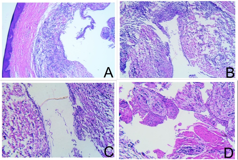
Presentation of case: A female patient presented with an asymptomatic gingival swelling in the lingual aspect of mandibular anterior region. The associated tooth (#34) was endodontically treated 3 years back. A periapical radiograph showed a well-defined round radiolucency on the tooth. Cone beam computed tomography (CBCT) revealed extensive bone destruction. The lesion was surgically excised and histological examination confirmed the diagnosis of LPC. The site healed satisfactorily post-operatively. The case was followed up for a year without any recurrence seen.
Discussion: LPC is a very rare clinical entity, the diagnosis of which requires a detailed case history taking, clinical and radiographic examination are essential to get proper assessment of the pathology. It is said to originate from either the remnants of dental lamina, reduced enamel epithelium or rests of Malassez. LPC presents with a typical histological picture which ensures the confirmatory diagnosis. Surgical enucleation with thorough curettage is the treatment of choice.
Conclusion: By reporting this rare case, we would like to stress to clinicians that there are a wide range of cysts and anatomic structures are present in the canine-premolar region of mandible of which LPC is a rare possibility.
Keywords: CBCT; Case report; Developmental cyst; Endodontically treated tooth; Gingivectomy; Lateral periodontal cyst.
Copyright © 2020 The Authors. Published by Elsevier Ltd.. All rights reserved.
Figures


Similar articles
-
Lateral periodontal cyst of the anterior maxilla: a rare case report.BMC Oral Health. 2025 May 7;25(1):690. doi: 10.1186/s12903-025-06044-9. BMC Oral Health. 2025. PMID: 40336026 Free PMC article.
-
Atypical presentation of lateral periodontal cyst associated with impacted teeth: two case reports.BMC Oral Health. 2021 Apr 7;21(1):178. doi: 10.1186/s12903-021-01539-7. BMC Oral Health. 2021. PMID: 33827538 Free PMC article.
-
The lateral periodontal cyst: aetiology, clinical significance and diagnosis.Endod Dent Traumatol. 2000 Aug;16(4):144-50. doi: 10.1034/j.1600-9657.2000.016004144.x. Endod Dent Traumatol. 2000. PMID: 11202873 Review.
-
Lateral periodontal cyst: a case report and literature review.J Oral Maxillofac Res. 2011 Jan 1;1(4):e5. doi: 10.5037/jomr.2010.1405. eCollection 2011. J Oral Maxillofac Res. 2011. PMID: 24421982 Free PMC article.
-
Lateral periodontal cyst. Review of the literature and report of a case.J Periodontol. 1990 Feb;61(2):126-31. doi: 10.1902/jop.1990.61.2.126. J Periodontol. 1990. PMID: 2179517 Review.
Cited by
-
Lateral periodontal cyst of the anterior maxilla: a rare case report.BMC Oral Health. 2025 May 7;25(1):690. doi: 10.1186/s12903-025-06044-9. BMC Oral Health. 2025. PMID: 40336026 Free PMC article.
References
-
- Shear M., Speight P.M. 4th edition. Blackwell Munksgaard; Oxford: 2007. Cysts of the Oral and Maxillofacial Regions; pp. 79–93.
-
- Friedrich R.E., Scheuer H.A., Zustin J. Lateral periodontal cyst. In Vivo (Athens, Greece) 2014;28(4):595–598. - PubMed
-
- Siponen M., Neville B.W., Damm D.D., Allen C.M. Multifocal Lateral periodontal cysts: a report of 4 cases and review of literature. Oral Surg. Oral Med. Oral Pathol. Oral Radiol. Endod. 2011;111(2):225–233. - PubMed
-
- Soluk-Tekkeşin M., Wright J.M. The World Health Organization classification of odontogenic lesions: a summary of changes of the 2017 (4th) edition. Turk. Patoloji Derg. 2018;34(1) - PubMed
Publication types
LinkOut - more resources
Full Text Sources
Research Materials
